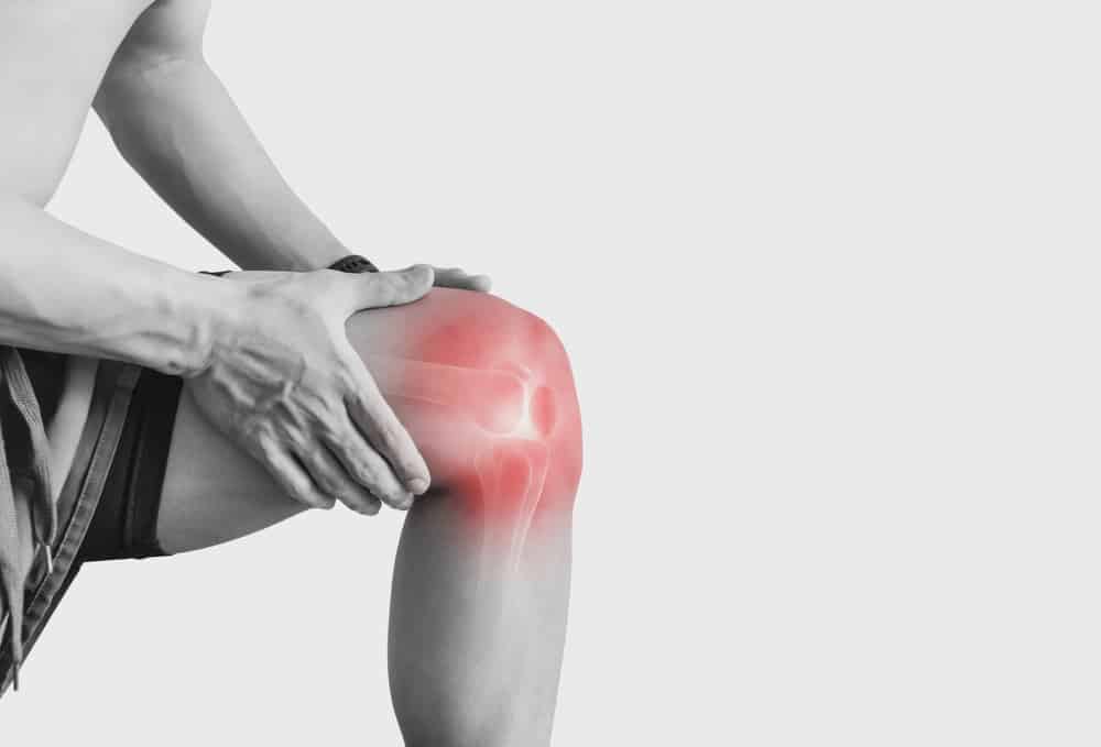

PCL injury diagnosis requires more than identifying pain after trauma. In many cases, the key clinical problem is posterior knee instability, which may be subtle in the acute setting and easier to underestimate than anterior laxity. A careful history, structured examination, and appropriately selected imaging help distinguish isolated injury from combined posterolateral or multiligament patterns. For clinicians, the goal is not only to confirm a posterior cruciate ligament injury, but also to grade instability, document functional impact, and identify associated lesions that alter management. This guide reviews practical examination steps, grading logic, imaging choices, and common pitfalls in daily practice.
1. PCL injury diagnosis starts with mechanism, symptoms, and suspicion
Early PCL injury diagnosis often depends on mechanism recognition. Classic mechanisms include a dashboard injury, a fall onto a flexed knee, or hyperflexion trauma in sport. Hyperextension injuries can also injure the PCL, especially when combined structures are involved.
Patients may not always describe dramatic instability. Instead, they may report posterior knee pain, swelling, difficulty decelerating, discomfort on stairs or declines, and a vague sense of mistrust during loaded flexion. Chronic presentations may feature anterior knee pain from altered tibiofemoral mechanics rather than an obvious complaint of giving way.
Because isolated PCL tears are less common than ACL injuries, under-recognition is a real issue. A useful starting point is a broad ligamentous laxity framework that separates symptoms, mechanism, static restraint injury, and functional instability.
In acute care or sports medicine settings, clinicians should also screen for combined injury patterns, neurovascular risk, major effusion, dislocation history, or inability to bear weight. A practical triage pathway may help prioritize urgent imaging and specialist follow-up when multiligament injury is suspected.
1.1 Clinical clues that raise suspicion
- Direct anterior tibial blow with the knee flexed
- Posterior proximal tibial tenderness or fullness
- Difficulty descending stairs or running downhill
- Perceived weakness in deceleration rather than overt buckling
- Possible associated PLC findings, especially if rotational symptoms are present
Knee laxity should be interpreted in context, because generalized laxity, guarding, pain, and swelling can affect exam findings.
2. PCL injury diagnosis on examination: what to test and how to avoid errors
The physical exam remains central to PCL injury diagnosis. The challenge is that a missed posterior tibial sag can distort every subsequent test. Before stressing the knee, inspect resting tibial position carefully.
2.1 Core examination maneuvers
The posterior sag sign is one of the most important early observations. With the patient supine and hips and knees flexed, compare the anterior tibial contour bilaterally. Loss of the normal anterior tibial step-off suggests posterior tibial translation at rest.
The posterior drawer test is the principal manual test for posterior translation. It is typically performed at 90 degrees of knee flexion, with the examiner ensuring neutral rotation and first reducing any posterior sag before applying a posteriorly directed force. This point matters: failure to reduce the tibia can underestimate the true endpoint and confuse grading.
The quadriceps active test may be helpful, especially when guarding limits the drawer test. Activation of the quadriceps from a flexed position can pull a posteriorly subluxed tibia anteriorly, supporting PCL insufficiency.
Rotational assessment is also important. The dial test knee helps detect associated posterolateral corner involvement, especially when asymmetry is more apparent at 30 degrees than at 90 degrees. This is not simply an added detail. Combined PCL-PLC injury changes both diagnosis and treatment planning.
For clinicians seeking a reproducible objective exam, documenting resting alignment, endpoint quality, side-to-side difference, and rotational findings in a consistent order improves serial comparison.
2.2 Common pitfalls in manual assessment
- Missing the resting posterior sag before the drawer test
- Not reducing the tibia before grading posterior translation
- Allowing hamstring contraction to resist posterior force
- Failing to compare with the contralateral knee
- Overlooking PLC injury when external rotation asymmetry is present
A broad overview of laxity testing can help standardize interpretation when multiple clinical maneuvers point in slightly different directions.
3. How to grade posterior instability clinically and structurally
Once injury is suspected, PCL injury diagnosis should progress to grading. In practice, clinicians are grading both ligament damage and the degree of posterior knee instability. These are related, but not identical, concepts.
PCL grading is often described using posterior tibial translation on examination or stress imaging. In broad clinical use, lower grades suggest partial or lower-magnitude posterior translation, while higher grades suggest complete injury or combined injury patterns. Exact thresholds can vary by method, examiner, and imaging protocol, so local consistency matters.
Isolated low-grade findings may be managed differently from high-grade posterior translation, recurvatum patterns, or combined PCL-PLC injury. That is why PCL injury diagnosis is not just yes-or-no confirmation.
3.1 A practical grading decision aid
- Confirm sag at rest before stressing the knee.
- Grade posterior translation on the posterior drawer test relative to the contralateral side.
- Check rotation with the dial test knee and varus alignment if PLC injury is possible.
- Correlate imaging if the exam is limited, equivocal, or suggests combined injury.
- Document function and symptoms, not only translation magnitude.
A useful comparison is the way clinicians approach valgus instability over time, where a structured grading workflow supports serial documentation. The same principle applies in posterior instability: use the same test conditions and language each time.
Recent reviews support this structured approach. Wilebski et al. (2026) discuss current standards for objective assessment and management of PCL tears, emphasizing the role of careful clinical and objective documentation. A concise management overview by Knapik et al. (2026) also reinforces that treatment decisions depend on instability grade, chronicity, symptoms, and associated injuries rather than MRI appearance alone.
4. Imaging in PCL injury diagnosis: MRI, stress radiographs, and when each helps
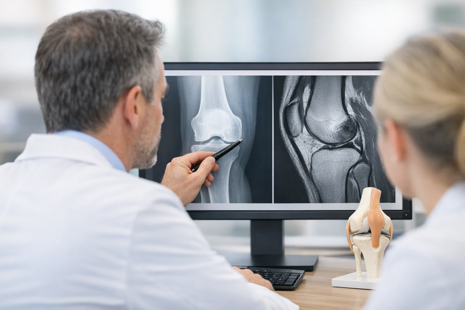
Imaging should answer different questions at different stages of PCL injury diagnosis. MRI is usually the best tool to visualize the ligament itself and to assess meniscal, chondral, osseous, and multiligament injury. However, MRI is a structural study, not a dynamic measure of posterior translation under load.
MRI posterior cruciate ligament assessment is particularly useful in acute injury, when clinicians need to characterize fiber continuity, edema, avulsion patterns, and associated damage. MRI remains complementary to clinical examination and is often important for preoperative planning when reconstruction is considered.
Stress imaging adds a different layer. Stress radiography PCL evaluation can quantify posterior tibial displacement and improve confidence in grading, especially in chronic or equivocal cases. This is valuable when the physical exam is limited by body habitus, pain, guarding, or examiner variability.
In a busy clinic, practical radiographic workflows matter. Lameire et al. (2026) describe a simple clinic-oriented approach to stress radiographs in multiligament injuries, reinforcing the usefulness of reproducible load application and side-to-side comparison. For isolated injury, Sirisopilkun et al. (2025) compared a modified Lachmeter technique with stress radiographic measurement in isolated PCL injury, highlighting the broader clinical need for objective posterior translation assessment.
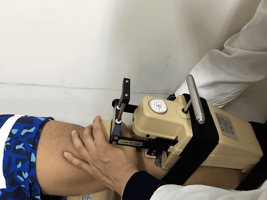
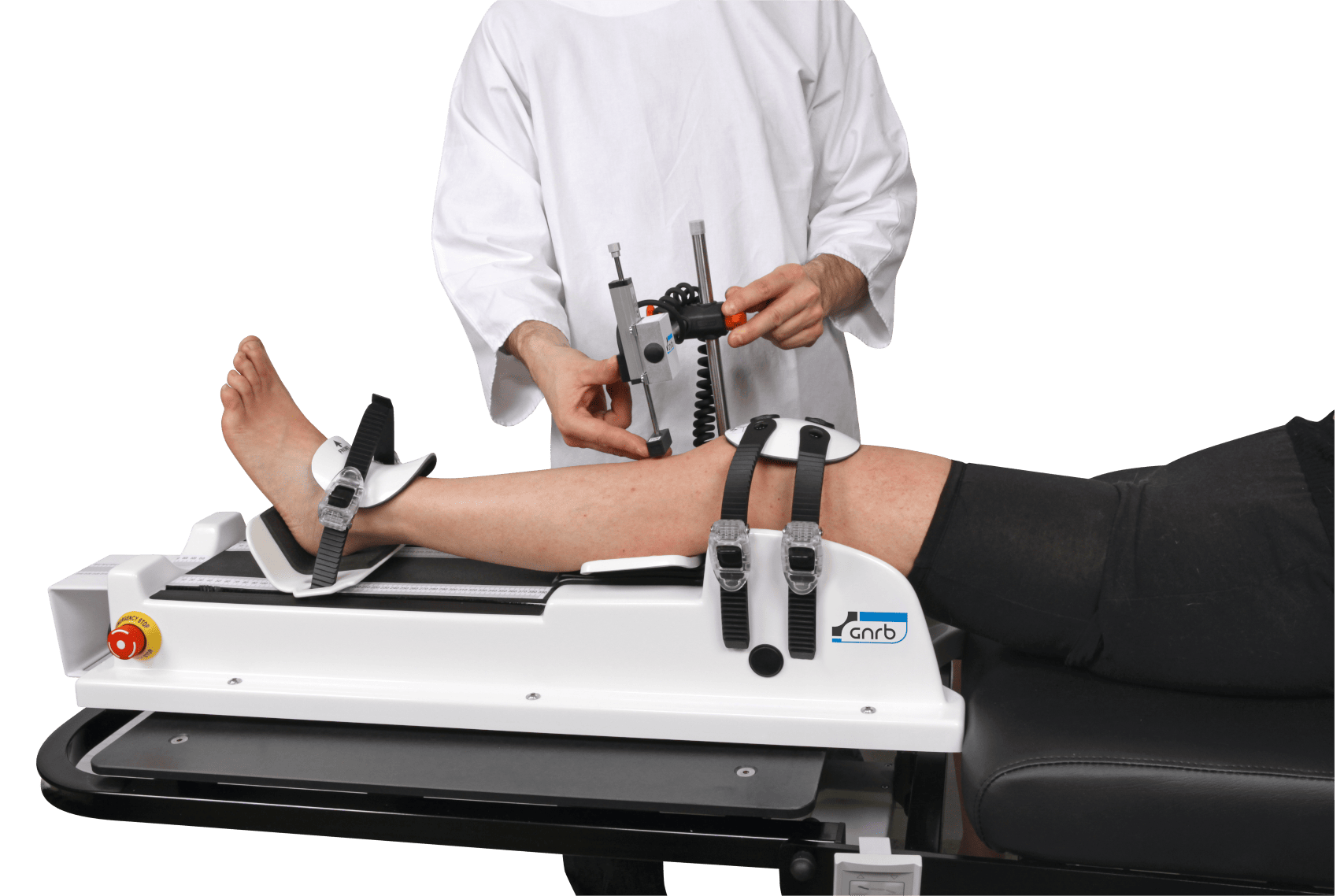
For clinicians interested in how loading devices and radiographic methods differ, this discussion of stress testing tools may help frame measurement consistency and practical constraints.
One more imaging point deserves attention: bone bruise distribution can support mechanism analysis in acute injury. Zou et al. (2025) examined bone contusion patterns in acute isolated PCL tears, which may help correlate MRI findings with the traumatic event and improve diagnostic confidence in selected cases.
5. Where objective laxity measurement may fit in posterior instability workup
When manual findings are subtle, serial monitoring is needed, or side-to-side comparison must be documented more precisely, objective assessment can strengthen PCL injury diagnosis by quantifying functional instability rather than relying only on descriptive endpoints. In posterior instability and combined injuries, a multi-axis workflow may be especially useful because posterior translation often coexists with coronal or rotational abnormalities.
These methods do not replace MRI. They may complement MRI and clinical examination by adding functional or dynamic information, helping clarify equivocal cases and improving follow-up documentation. General principles of dynamic testing and arthrometer evaluation are relevant when a clinic aims to standardize instability measurement across time or across examiners.
6. Putting it together: a clinic-ready pathway for PCL injury diagnosis
In real practice, PCL injury diagnosis works best as a layered process. History suggests mechanism. Examination identifies resting sag and posterior translation. Rotational testing checks for associated PLC injury. MRI defines structural damage and associated lesions. Stress or objective testing may refine grading and follow-up when needed.
Suggested workflow
- Start with mechanism, swelling, and functional complaint.
- Inspect for posterior sag before any stress test.
- Perform the posterior drawer test, quadriceps active test, and dial test knee.
- Order MRI posterior cruciate ligament evaluation when structural definition or associated injury assessment is needed.
- Consider stress radiography PCL assessment when grading remains uncertain or objective posterior translation documentation would influence decisions.
- Reassess over time, especially in chronic or combined injury patterns.
This structured approach supports clearer communication among orthopaedic surgeons, sports physicians, physiotherapists, and radiologists. It also improves follow-up because repeated measurements are only clinically meaningful if the initial PCL injury diagnosis was documented in a reproducible way.
PCL injury diagnosis should remain clinician-led and individualized. Exam findings, symptoms, activity demands, chronicity, and associated pathology all shape next steps. PCL injury diagnosis is strongest when no single test is overvalued in isolation. A posterior drawer test without sag assessment can mislead. MRI without functional grading can miss the practical severity of instability. Stress measures without clinical context can also be misread.
7. Key takeaways and next steps
PCL injury diagnosis depends on combining mechanism, examination, and imaging rather than relying on one element alone. The highest-yield bedside findings remain the posterior sag sign and posterior drawer test, with the dial test knee helping identify associated posterolateral injury. PCL grading should reflect true posterior instability, not just an imaging label.
MRI is essential for structural assessment and associated injury evaluation, while stress radiography PCL methods or other objective measurements may help quantify side-to-side instability in selected cases. For teams managing these injuries longitudinally, the next step is usually to standardize documentation, align grading terminology, and match treatment decisions to instability pattern, symptoms, and activity demands.
If uncertainty remains after initial assessment, repeat examination once swelling and guarding improve, and correlate findings across modalities rather than forcing a decision from a single imperfect test. That is often the most reliable path to a confident PCL injury diagnosis.
Clinical references (PubMed)
1) 2026 – Wilebski et al. – Current standards for the objective assessment and management of posterior cruciate ligament tears: a narrative review. – Annals of Joint – DOI: 10.21037/aoj-25-52 – PMID: 41657676 – PubMed
2) 2025 – Sirisopilkun et al. – Comparison Between the Modified Lachmeter Technique and Stress Radiographic Measurement in an Isolated Posterior Cruciate Ligament Injury. – Orthopaedic Journal of Sports Medicine – DOI: 10.1177/23259671251391352 – PMID: 41268057 – PubMed
3) 2026 – Lameire et al. – Assessment of Laxity in Multiligamentous Knee Injuries Using Stress Radiographs: A Simple Technique for a Busy Clinic. – Video Journal of Sports Medicine – DOI: 10.1177/26350254251378514 – PMID: 41834844 – PubMed
4) 2026 – Knapik et al. – Management of Posterior Cruciate Ligament Injury: A Concise Overview of Current Indications, Techniques, and Outcomes. – Journal of the American Academy of Orthopaedic Surgeons – DOI: 10.5435/JAAOS-D-25-00500 – PMID: 41202194 – PubMed
5) 2025 – Zou et al. – Distribution of Bone Contusion Patterns in Knees with Acute Isolated Posterior Cruciate Ligament Tears. – Orthopaedic Journal of Sports Medicine – DOI: 10.1177/23259671251397639 – PMID: 41409449 – PubMed