

Study Title: Which tibial implantation site for the deep medial collateral ligament should be chosen to control anteromedial rotatory instability of the knee?
Authors: Antoine Hamon1 | Harold Common1 | Théo Cojean | Henri Robert
Journal: Journal of Experimental Orthopaedics
Publication Date: 2025
DOI: 10.1002/jeo2.70585
Institution: Orthopaedic Department, University Hospital, Rennes, France
University Claude Bernard, Lyon 1, France
Orthopaedic Department, Hospital Haut Anjou, Château‐Gontier‐sur‐Mayenne, France
This study highlight reviews a 2025 cadaveric paper that explored whether changing the tibial insertion site of an anteromedial reconstruction strand could improve control of anteromedial rotatory instability in severe medial knee injury. The authors focused on Deep MCL reconstruction within a broader trifid flat graft construct designed to reproduce the superficial MCL, deep MCL, and posterior oblique ligament. Their question is clinically relevant because conventional medial reconstructions do not always restore native rotational stability, especially when deep medial structures and the anteromedial capsule contribute to persistent laxity.
Why this study matters
Medial knee injuries are often more complex than they first appear, especially when they occur with ACL rupture. The paper reviews prior work showing that injuries to the superficial MCL, deep MCL, posteromedial structures, and anteromedial capsule may all influence rotational control. In that setting, Deep MCL reconstruction is not just about restoring valgus restraint. It may also help address external rotation and anteromedial rotatory instability that can remain after more traditional techniques.
The authors specifically questioned whether the tibial insertion of the anteromedial strand should remain close to the classic anatomic orientation or whether a more oblique, more isometric course might better control laxity.
Study design and what was tested
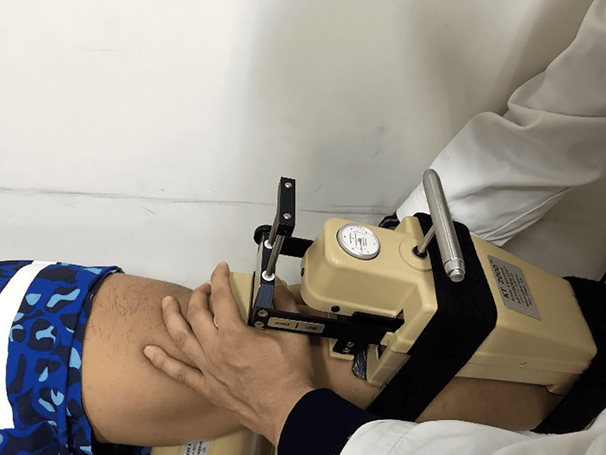
This was a Level V experimental cadaveric study using 20 fresh-frozen knees from 13 female and 7 male donors with a mean age of 80 years. The team tested the knees at 30 degrees of flexion using the Dyneelax static laximeter.
After baseline measurements in the intact state, the authors performed standardized transections of the superficial MCL, deep MCL, and posterior oblique ligament. They then reconstructed the medial side using a trifid flat graft with three components: an anterior strand for the deep layer and anteromedial function, an intermediate strand for the superficial MCL, and a posterior strand for the posterior oblique ligament.
Two tibial positions for the anterior or anteromedial strand were compared:
- An anatomical-style position with an angle alpha of 20 degrees or less relative to the superficial MCL
- A more oblique isometric position with an angle alpha greater than 20 degrees
In practical terms, the difference was the amount of anteriorization of the tibial insertion. This made the Deep MCL reconstruction either relatively less oblique or more oblique.
What outcomes were measured
The study measured three variables at 30 degrees of flexion:
- Anterior tibial translation under 200 N
- Internal rotation under 5 N-m
- External rotation under 5 N-m
The authors reported both absolute laxity values and residual laxity after reconstruction compared with the intact knee. This is important because the central question was not simply whether reconstruction helped, but whether one tibial position for Deep MCL reconstruction restored stability more effectively than the other.
Key findings
Both reconstruction strategies significantly reduced laxity compared with the transected state. That means either version of the trifid flat graft may help recover some stability after a complete medial injury pattern.
However, the more oblique configuration, defined as alpha greater than 20 degrees, performed better in two important areas. Residual anterior tibial translation was lower in the more oblique group than in the less oblique group: 0.74 +/- 0.58 mm versus 1.30 +/- 0.56 mm, with p = 0.04. Residual external rotation was also lower: 0.35 +/- 0.39 degrees versus 1.05 +/- 0.89 degrees, also with p = 0.04.
For internal rotation, the paper reports no significant difference between groups in the abstract, with p = 0.24. In the main results tables, reconstruction values for internal rotation are presented and should be interpreted with caution, but the overall message remains that the clearer advantage of this Deep MCL reconstruction strategy appeared in anterior translation and external rotation rather than internal rotation.
The authors also note that sectioning created similar baseline instability in both groups before reconstruction, which supports the view that the post-reconstruction differences were related to the tibial insertion strategy rather than unequal injury effects.
How the authors interpreted the biomechanical effect
The paper suggests that a more oblique anterior strand may create a stronger horizontal restraining vector. In simple terms, as the angle between the anteromedial strand and the superficial MCL increases, the graft may do more to resist anterior shear and external rotation. This may explain why the more oblique version of Deep MCL reconstruction appeared to better control anteromedial rotatory laxity.
The study also frames this within the broader understanding of medial knee anatomy. The deep MCL and anteromedial capsule are increasingly recognized as important restraints to external rotation near extension and through early flexion. A reconstruction that better reproduces this oblique functional orientation may therefore be more effective than one that is only anatomically close in footprint but mechanically less favorable.
Why the flat trifid graft concept is relevant
This paper was not only about tunnel position. It also supports the idea that broad, flat medial structures may not be fully replicated by round grafts alone. The authors used a trifid flat graft to mimic the superficial MCL, the deep layer, and the posterior oblique ligament in a more tissue-like configuration.
That does not prove that every flat construct is superior in every patient, but it fits with recent biomechanical work suggesting that flat graft geometry may distribute load more naturally across knee motion. In this context, Deep MCL reconstruction was part of a more comprehensive attempt to restore native medial restraint patterns rather than simply reconstruct one cord-like bundle.
Where objective laxity testing fits
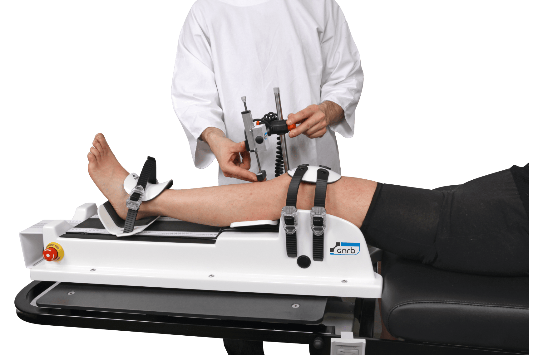
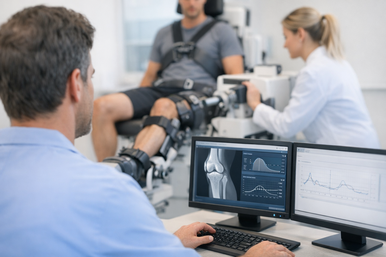
This study is a good example of why objective knee laxity data can be useful in ligament research and follow-up. The authors did not rely only on visual assessment or manual testing. They used the Dyneelax device to quantify anterior translation and tibial rotation under standardized loads, which helped detect the differences between the two reconstruction strategies.
In clinical practice, that type of objective information may support serial assessment after medial-sided surgery, especially when the question is whether a patient still shows measurable residual laxity despite reconstruction. Devices such as the Dyneelax knee arthrometer may help document translational and rotational behavior under controlled conditions, while the GNRB arthrometer may support objective anterior laxity assessment in ACL-related pathways.
Still, these tools should be viewed as complementary. They can add reproducible data to the clinical exam and may help with follow-up or functional comparison, but they do not replace MRI for defining the injured structures, graft position, associated meniscal damage, or other intra-articular pathology. For a case involving possible Deep MCL reconstruction, imaging, examination, and patient-specific context remain essential.
Limits to keep in mind
The authors are appropriately cautious. This was a cadaveric study, not a clinical outcomes trial. The sample size was 20 knees, and the donors were elderly, with a mean age of 80 years. Bone quality and tissue properties in this setting may differ substantially from younger athletic patients.
Testing was performed only at 30 degrees of flexion because of the device setup, and valgus testing was not possible with this protocol. Graft tensioning was manual rather than standardized with a force gauge, which may introduce variability. The ACL was left intact, so the findings isolate the medial reconstruction question but do not fully reproduce combined injury patterns commonly seen in practice.
For all of these reasons, the study suggests a potentially better tibial insertion concept for Deep MCL reconstruction, but it does not establish definitive surgical superiority in living patients.
Clinical interpretation
For surgeons treating severe medial instability with anteromedial rotatory features, this study may support considering a more oblique tibial insertion for the anterior strand within a combined medial reconstruction. The likely benefit appears to be better control of external rotation and some improvement in anterior translation. That said, residual laxity remained in both groups, and the authors themselves describe the result as a trend that requires confirmation.
So the most balanced take is this: Deep MCL reconstruction may be more effective biomechanically when the anterior strand is placed in a more oblique orientation, but clinical validation is still needed before broad conclusions are made.
Key takeaways
- This cadaveric study suggests that a more oblique tibial insertion for the anteromedial strand may improve Deep MCL reconstruction control of anterior translation and external rotation.
- Both reconstruction strategies reduced laxity after complete medial transection, but neither fully restored native stability.
- The work supports the idea that medial knee reconstruction may need to address deep and anteromedial structures, not only the superficial MCL and posterior oblique ligament.
- Objective laxity testing can help quantify subtle differences in reconstruction behavior, but it complements rather than replaces MRI and expert clinical examination.
- Because this was an elderly cadaveric model tested at one flexion angle, the findings should be interpreted cautiously until supported by clinical studies.