
anteromedial instability knee is a clinically important but often under-recognized pattern of medial knee instability that sits at the intersection of valgus laxity, anterior tibial translation, and internal-external rotational control. In practice, it rarely reflects failure of one structure alone. Instead, the superficial and deep medial restraints, posterior structures, cruciate status, and even the meniscus may all influence presentation. For orthopaedic surgeons, sports physicians, physiotherapists, and researchers, the challenge is not only naming the lesion, but understanding which medial structures matter most, how to examine them, and when combined injury patterns should change management.
1. anteromedial instability knee in modern clinical terms
The term anteromedial instability knee generally refers to abnormal anterior and rotational subluxation behavior of the medial tibial plateau relative to the femur, usually associated with deficiency within the medial knee ligament complex and often influenced by cruciate injury status. Although the phrase is sometimes used interchangeably with anteromedial rotatory instability, the latter more explicitly highlights the rotational component.
Clinically, anteromedial instability knee is best thought of as a spectrum rather than a single diagnosis. Some patients present with obvious valgus laxity after trauma. Others report persistent giving way after ACL reconstruction, equivocal medial pain, or instability that seems disproportionate to MRI findings. This is one reason why understanding the medial plane is so important when evaluating recurrent or combined instability.
The relevant restraints are not limited to the superficial MCL. The medial knee ligament complex includes the superficial medial collateral ligament, deep MCL components, posterior oblique ligament, capsule, and dynamic muscular influences. Depending on knee flexion angle and load direction, different tissues dominate resistance to valgus and rotation. That complexity explains why isolated labels can be misleading in both diagnosis and surgical planning.
2. Medial restraints that shape anteromedial instability knee
2.1 Superficial and deep layers
The superficial medial collateral ligament remains the principal static restraint to valgus stress, especially through mid-range flexion. It also contributes to rotational control, particularly when injury extends beyond a simple proximal or midsubstance lesion. However, the deep layer has gained increasing attention in discussions of anteromedial instability knee because of its role in controlling pathologic tibial motion close to the joint line.
Recent biomechanical work has reinforced this concept. Behrendt et al. (2025) reported that anteromedial reconstructions that mimic deep MCL function can reduce ACL force, supporting the idea that medial deficiency may amplify cruciate loading rather than simply coexist with it. This has direct implications for revision cases, residual pivot-related symptoms, and combined injury strategies.
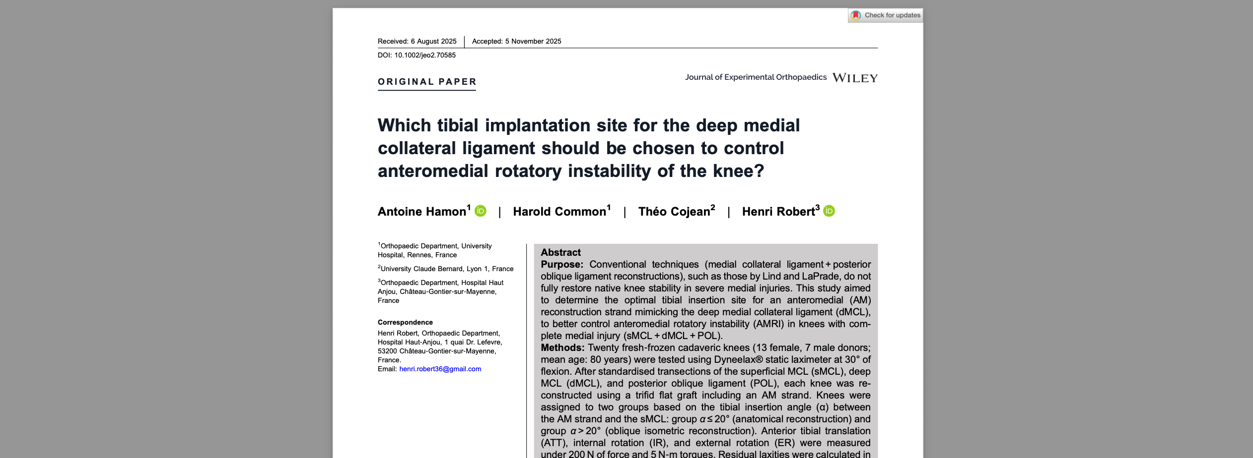
For surgeons focused on insertional anatomy and rotatory control, the question of where the deep MCL is tensioned and fixed is central to restoring anteromedial instability knee. The article on deep MCL insertion concepts is particularly relevant when trying to connect anatomy with observed laxity patterns.
2.2 Posterior support and the AM-PM concept
The anteromedial and posteromedial functional concepts are useful because they remind clinicians that the medial side is not uniform. The posterior oblique ligament contributes importantly to posteromedial and rotational control, especially near extension, and often becomes clinically relevant when grade III injuries or combined lesions are present.
A 2026 systematic review by Ade-Conde et al. (2026) found favorable patient-reported outcomes and medial stability after combined MCL and posterior oblique ligament reconstruction in grade III injuries. While this does not mean every patient with anteromedial instability knee needs dual reconstruction, it supports the broader message that persistent instability may reflect failure to address the full medial envelope.
When planning reconstruction, it can also help to compare different reconstruction philosophies through the lens of translation and rotation. The discussion of reconstruction concepts is useful for linking biomechanical goals to operative decision-making.
2.3 Meniscal contribution
The medial meniscus restraint should not be ignored. In some knees, meniscocapsular or meniscal root-related dysfunction may alter secondary restraint behavior and magnify symptoms that resemble or compound anteromedial instability knee. Meniscal pathology alone does not define anteromedial rotatory instability, but it may reduce the knee’s ability to buffer translation and load sharing.
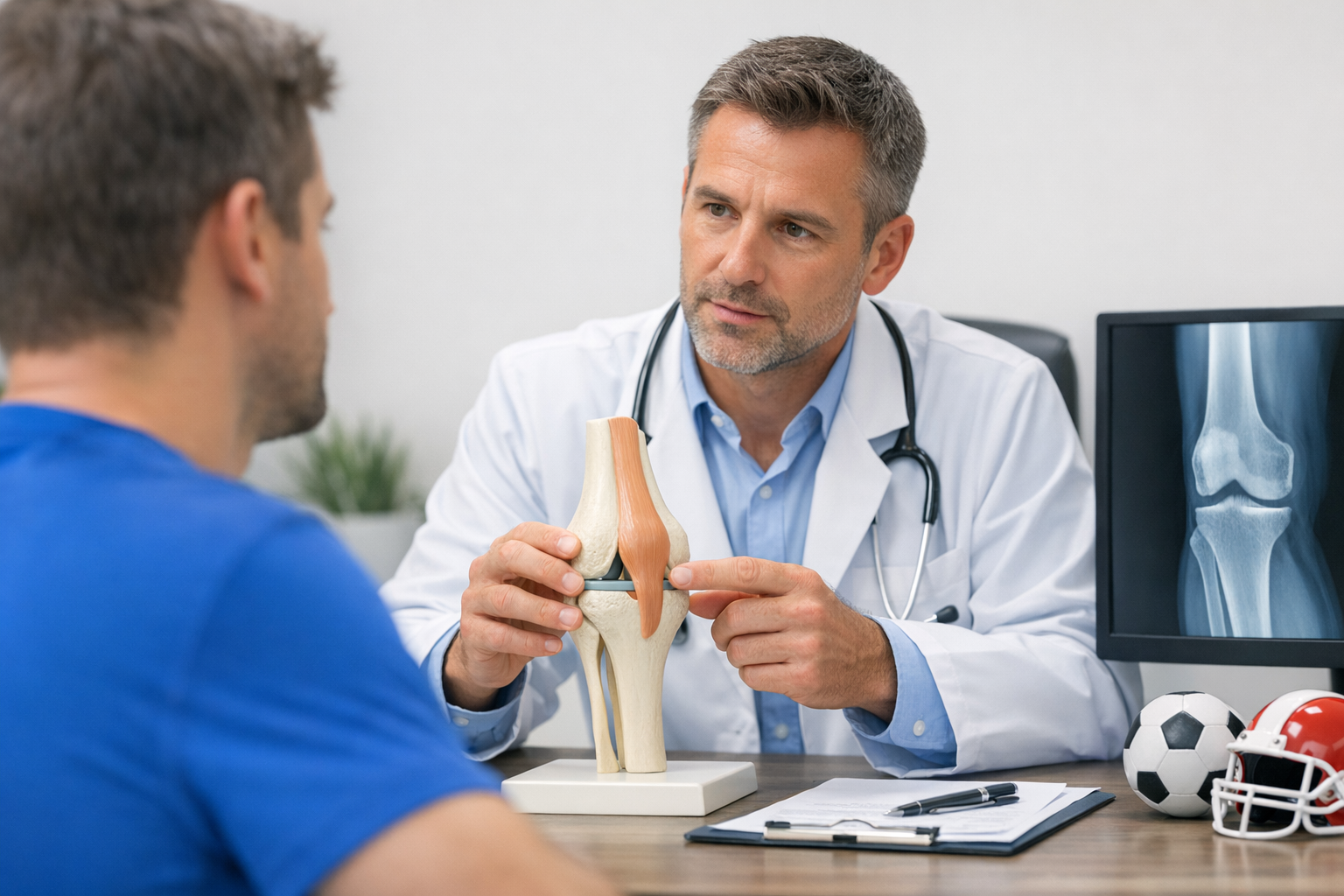
This is one reason MRI remains important even when instability is strongly suspected clinically. Functional testing can describe how unstable the knee behaves, but MRI is still typically needed to evaluate meniscal, chondral, osseous, and associated ligament injury patterns, especially when surgery is under consideration.
3. How anteromedial instability knee presents in clinic
Patients with anteromedial instability knee may describe a vague sense of the knee shifting inward, collapsing with cutting or deceleration, or feeling unstable during pivoting and downhill activity. Some report instability after a known MCL injury that never fully normalized. Others present after ACL injury or reconstruction with residual symptoms that are not fully explained by sagittal testing alone.
Typical clues include:
- Medial joint line or proximal/distal MCL tenderness
- Perceived valgus opening or inward collapse
- Instability near extension or during directional change
- Persistent symptoms despite apparently healed isolated MCL injury
- Combined cruciate findings, especially in complex trauma
Because valgus rotational instability can be subtle, clinicians should be cautious not to over-rely on a single maneuver. MRI may under-represent the functional severity of rotational patterns, as discussed in rotational instability. That does not diminish the value of MRI. It simply reinforces that image appearance and real-world instability are not always equivalent.
In multiligament trauma, anteromedial instability knee may be only one component of a broader picture. If the injury mechanism, bruising pattern, or examination suggests combined pathology, a structured checklist approach helps reduce missed lesions.
4. Clinical examination for anteromedial instability knee
A good clinical examination medial knee starts with comparison. Side-to-side asymmetry, endpoint quality, flexion angle dependence, and symptom reproduction all matter. The question is not only whether valgus opens the joint, but whether the pattern suggests isolated superficial MCL injury, deeper meniscocapsular involvement, posterior oblique deficiency, cruciate interaction, or a combined lesion producing anteromedial instability knee.
4.1 Core examination maneuvers
Valgus stress testing at 30 degrees remains foundational for assessing the superficial MCL, while testing near full extension helps screen for broader medial or cruciate involvement. A structured valgus workflow can improve grading consistency and follow-up interpretation.
Additional exam points may include:
- Valgus stress at 0 and 30 degrees
- Assessment of anterior translation and endpoint quality
- Rotational comparison during pivot-related maneuvers
- Palpation of femoral, midsubstance, and distal medial structures
- Evaluation for meniscal irritation, locking, or joint line tenderness
No single test fully defines anteromedial instability knee. Instead, clinicians synthesize the injury mechanism, location of pain, valgus opening, rotational behavior, and associated ligament findings. A broad review of mechanics can help frame these findings in a more reproducible way.
4.2 A short decision aid
When anteromedial instability knee is suspected, this practical sequence can help:
- Confirm the symptom pattern – valgus collapse, rotational giving way, extension-related instability, or recurrent medial-sided symptoms.
- Localize tenderness – superficial MCL, distal deep structures, posteromedial corner, or meniscocapsular zone.
- Test at multiple angles – especially 0 and 30 degrees, noting whether laxity is isolated or combined.
- Check cruciate interaction – particularly when symptoms exceed apparent isolated MCL findings.
- Correlate with imaging – MRI for associated injury assessment and operative planning where needed.
The article on combined ACL-MCL injury is especially useful when anterior translation and medial deficiency coexist, since that scenario often raises the question of whether anteromedial instability knee is contributing to residual laxity.
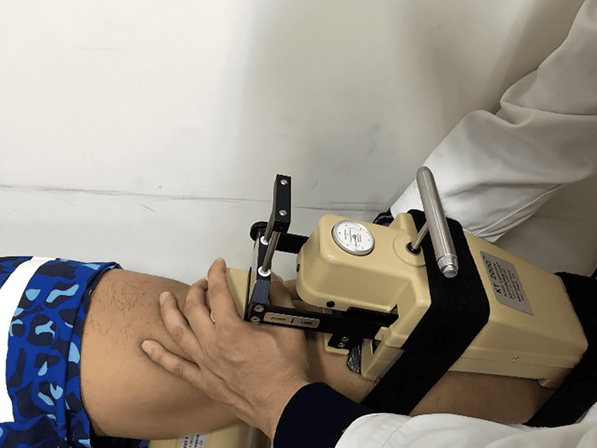
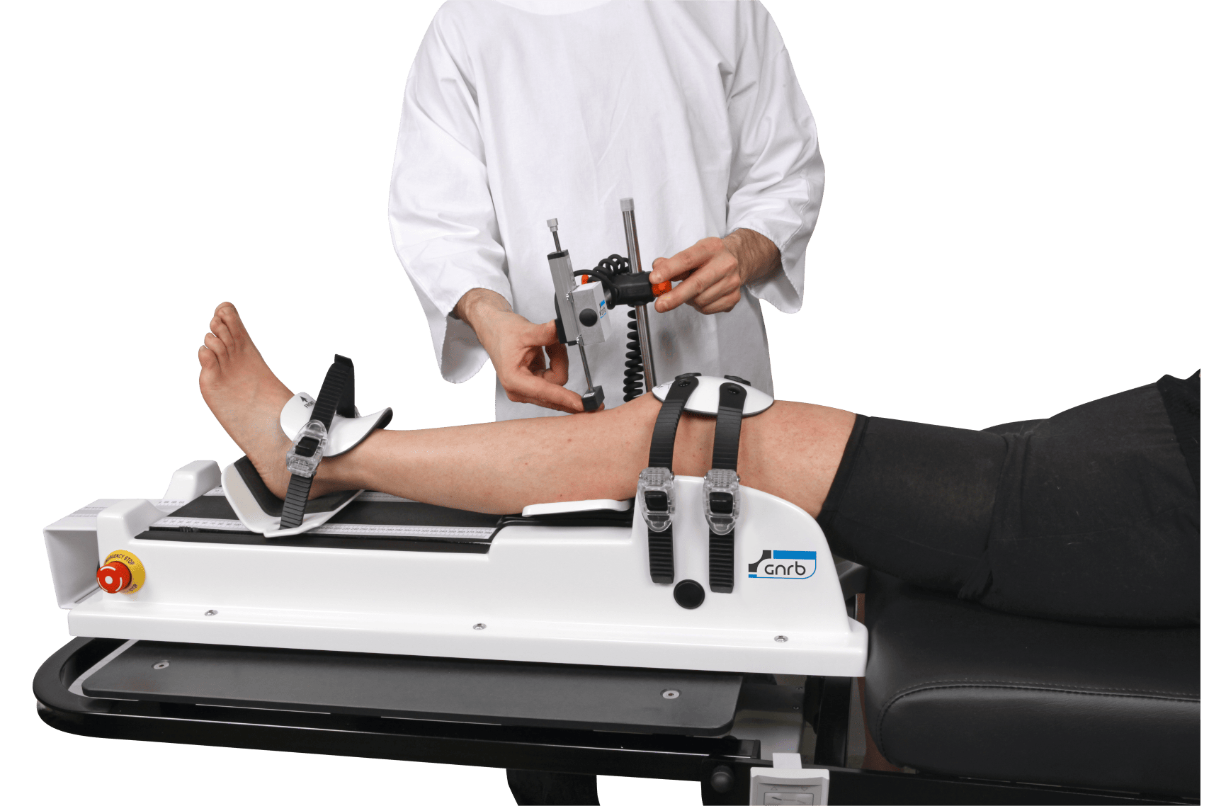
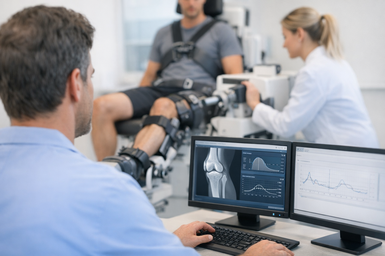
One sentence on objective testing is warranted here: in selected cases of suspected complex or borderline medial-sided laxity, quantified multi-axis assessment with devices such as the GNRB arthrometer or Dyneelax may complement MRI and hands-on examination by adding functional side-to-side data rather than replacing imaging.
5. Interpreting laxity patterns and combined injury mechanisms
The main pitfall in anteromedial instability knee is oversimplification. Medial opening does not always equal isolated superficial MCL injury, and residual anterior instability after ACL reconstruction does not always indicate graft failure alone. The medial side can increase translational and rotational demand on the ACL, and the ACL can mask or magnify medial pathology depending on injury timing and examination conditions.
This relationship is reflected in emerging biomechanical evidence. Gellhaus et al. (2025) biomechanically validated different anteromedial reconstruction techniques, emphasizing that reconstruction strategy influences how effectively instability is controlled. Taken together with Behrendt et al. (2025), the message is that anteromedial instability knee is not just a descriptive term. It has mechanical consequences that may affect cruciate loading and operative planning.
For clinicians using quantified assessment pathways, objective examination may help document instability over time, especially when manual grading feels equivocal. In more complex cases, a multi-axis assessment approach is often more aligned with the actual problem than a single-plane measure.
Important interpretation points include:
- Laxity angle matters – opening only at 30 degrees suggests a different pattern from opening in near extension.
- Rotation matters – pure valgus and combined valgus-rotational behavior should not be treated as identical.
- Associated restraints matter – ACL, POL, deep MCL, capsule, and meniscus may all alter the exam.
- Symptoms matter – some patients have measurable laxity but limited functional instability, while others are highly symptomatic with less dramatic manual findings.
6. Management concepts and practical takeaways for clinicians
Treatment of anteromedial instability knee depends on acuity, severity, tissue quality, associated injuries, and patient demands. Not every case requires surgery, and not every medial injury with some rotational features should be labeled a major reconstructive problem. Still, clinicians should maintain a low threshold for reassessing persistent instability, especially after high-grade injury, combined cruciate trauma, or ongoing symptoms after rehabilitation.
Recent literature provides useful signals without justifying overstatement. Azuma et al. (2026) described semimembranosus transposition as an augmentation technique for anteromedial rotatory instability in a retrospective case series, while Thürig et al. (2026) presented a modified refixation technique for distal deep and superficial MCL injuries. These reports support continued refinement of medial-sided procedures, but diagnosis and treatment should remain clinician-led and individualized.
Key takeaways and next steps
- anteromedial instability knee is usually a combined functional problem, not a single-structure label.
- The superficial medial collateral ligament, deep MCL, and posterior oblique ligament all deserve attention in clinical reasoning.
- Medial knee instability should be assessed across valgus, translation, and rotation, not in one plane only.
- MRI remains complementary and important for associated injuries and surgical planning.
- Persistent symptoms after MCL or ACL injury should prompt reconsideration of unrecognized anteromedial instability knee.
In daily practice, the most useful approach is disciplined pattern recognition: define the symptoms, localize the injured tissues, examine at multiple angles, correlate with imaging, and reassess function over time. That framework is often more valuable than trying to force every patient into an isolated ligament label.
Clinical references (PubMed)
1) 2025 – Gellhaus et al. – Optimizing the Control of Anteromedial Rotatory Knee Instability: A Biomechanical Validation of Different Anteromedial Reconstruction Techniques. – Am J Sports Med – DOI: 10.1177/03635465251339820 – PMID: 40371817 – PubMed
2) 2025 – Behrendt et al. – Anterior Cruciate Ligament Force Is Reduced by Anteromedial Reconstructions That Mimic the Role of Deep Medial Collateral Ligament. – Am J Sports Med – DOI: 10.1177/03635465251387280 – PMID: 41170921 – PubMed
3) 2026 – Azuma et al. – Transposition of the Semimembranosus as an Augmentation Technique for Anteromedial Rotatory Instability of the Knee: A Retrospective Case Series Study. – J Knee Surg – DOI: 10.1055/a-2779-0226 – PMID: 41529724 – PubMed
4) 2026 – Ade-Conde et al. – Combined medial collateral ligament and posterior oblique ligament reconstruction demonstrates favourable patient-reported outcomes and medial knee stability in Grade III injuries: A systematic review. – Knee Surg Sports Traumatol Arthrosc – DOI: 10.1002/ksa.70344 – PMID: 41711578 – PubMed
5) 2026 – Thürig et al. – Modified Refixation Technique of Distal Deep and Superficial Medial Collateral Ligament Injuries. – Arthrosc Tech – DOI: 10.1002/atn2.70016 – PMID: 42382921 – PubMed