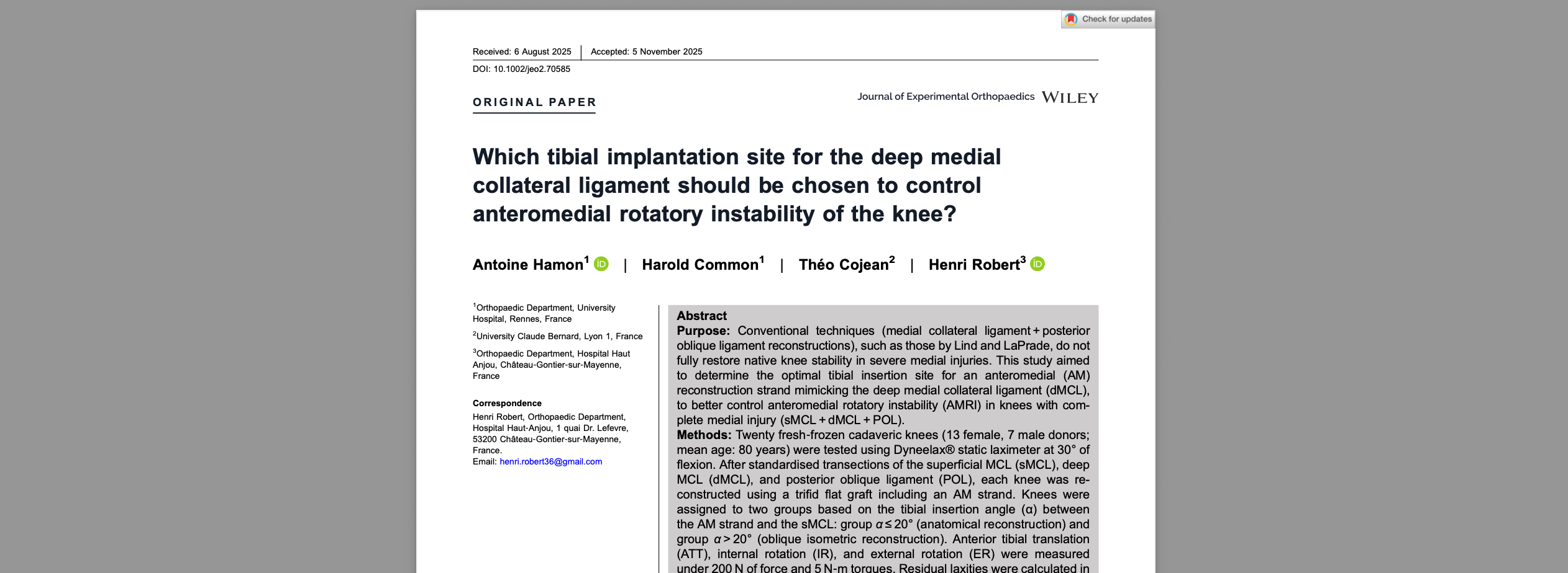
From image to function: why anatomy alone is not enough
Modern knee stability assessment cannot rely on anatomy in isolation. MRI can define fiber continuity, edema, graft appearance, bone bruising, meniscal tears, and cartilage injury, but it does not always explain why a patient still reports giving way, poor confidence on cutting, or an abnormal pivoting sensation. That gap is where functional knee instability becomes clinically important. In daily practice, surgeons, sports physicians, physiotherapists, and radiologists increasingly need a combined model that links morphology to mechanics, especially for ACL injury, rotational instability, and post-reconstruction follow-up.
A robust knee stability assessment therefore combines clinical examination, imaging, and objective knee laxity testing. This is not about replacing MRI. It is about adding measured function to structural interpretation, so that symptoms, manual testing, and imaging findings can be read in the same clinical frame.
1. Why knee stability assessment needs both anatomy and mechanics
The knee is stable because of an interaction between passive restraints, active neuromuscular control, and task-specific loading. The ACL, PCL, MCL, LCL, capsule, menisci, and anterolateral structures all contribute, but the way they contribute varies with flexion angle, axial rotation, quadriceps-hamstring co-contraction, gait speed, landing, and pivoting demands.
This matters because a structurally visible ligament is not always a functionally competent ligament. Likewise, a graft that appears acceptable on imaging may still allow measurable translational or rotational excess under load. A complete knee stability assessment should therefore ask four linked questions:
- What is injured or reconstructed anatomically?
- How much side-to-side laxity exists objectively?
- Is instability mainly translational, rotational, or combined?
- Does the measured mechanics match the patient’s symptoms and function?
That framework is especially useful in ACL-deficient knees, partial tears, graft surveillance, revision work-up, and suspected residual pivot shift. A useful overview of this integrated model is outlined in an objective examination framework that brings anatomy and biomechanics into the same visit.
In other words, MRI shows what the knee looks like. Mechanics help show what the knee does.
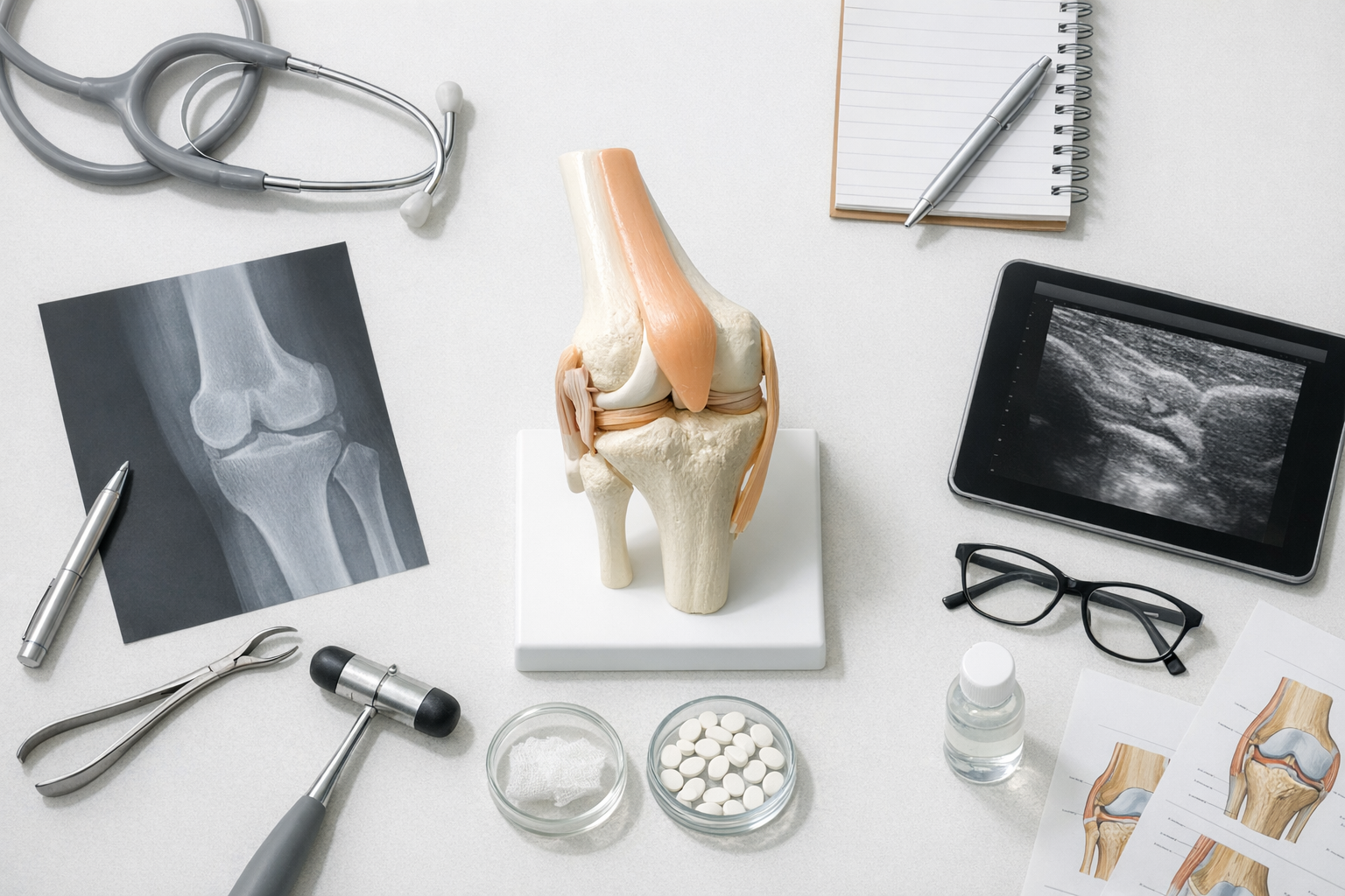
2. MRI is essential, but MRI and knee mechanics answer different questions
MRI remains central to the diagnostic pathway. It identifies associated meniscal tears, chondral lesions, bone bruising, tunnel issues, graft signal characteristics, and soft-tissue injury patterns that matter for treatment planning. For many referrals, MRI is indispensable. Still, MRI and knee mechanics should be viewed as complementary, not competing.
This distinction is becoming more important in the recent literature. Figueroa et al. (2025) examined the relationship between quantitative MRI UTE T2* findings in ACL autografts and BMI-normalized knee laxity within the first postoperative year. The clinical implication is not that imaging is less valuable, but that graft tissue characteristics and measured laxity may inform different dimensions of recovery. A graft can look maturing on MRI while function still evolves under load.
Similarly, Moretti et al. (2025) explored anterior knee laxity and ACL MRI signals in healthy and reconstructed knees following exercise. This kind of work reinforces a practical point for knee stability assessment: image interpretation may be influenced by timing and context, while laxity measurement can add a direct mechanical readout.
In nonoperative pathways, the mismatch can be even more clinically relevant. Dayal et al. (2026) systematically reviewed patients with ACL tears treated without surgery and reported that many showed persistent laxity on arthrometer assessment despite MRI evidence of fiber continuity. This does not mean MRI is wrong. It means visible continuity does not automatically equal functional competence.
That is particularly relevant in borderline MRI cases, where MRI remains necessary for associated injury assessment and surgical planning, but quantitative instability testing may help clarify whether a symptomatic knee behaves like a stable one.
For clinicians facing equivocal imaging, a balanced pathway is to pair MRI interpretation with side-to-side measurement and examination findings rather than over-weight any single test. The relationship between structure and function is also discussed in this article on MRI versus arthrometer use in ACL pathways.
3. What objective knee laxity testing adds to knee stability assessment
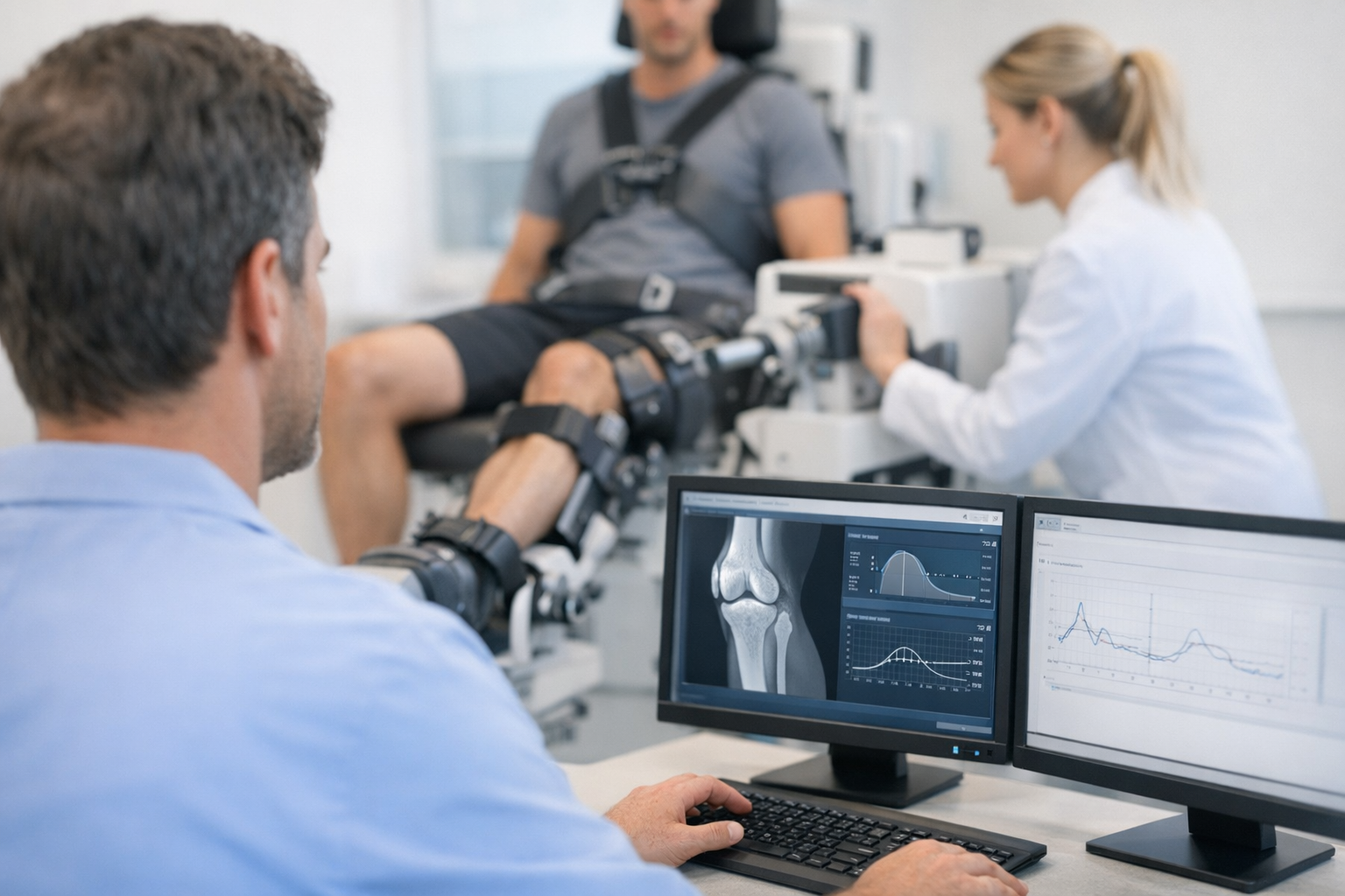
Objective knee laxity testing gives a quantified estimate of mechanical behavior under standardized load. In ACL work-up, this often means anterior tibial translation and side-to-side difference. More advanced systems can also examine compliance, stiffness, or multi-axis behavior relevant to dynamic knee stability and rotational control.
The practical value is straightforward. Manual tests remain essential, but they are examiner-dependent and can be influenced by guarding, swelling, anatomy, and experience. An instrumented knee arthrometer can help standardize the signal, especially when symptoms, MRI, and bedside findings do not fully align.
3.1 When measured mechanics are especially useful
A more complete knee stability assessment may benefit from instrumented testing in the following situations:
- Suspected partial ACL tear with persistent instability symptoms
- Equivocal Lachman or guarded examination
- Post-ACL reconstruction follow-up
- Return-to-sport decision support
- Revision ACL evaluation
- Residual pivot shift or suspected combined injury pattern
For ACL-specific pathways, this is where ACL laxity measurement becomes clinically meaningful. Side-to-side difference is useful, but it is not the only signal. Mechanical behavior under progressive load, endpoint quality, and load-displacement pattern may provide additional context. That principle is explored further in this discussion of why compliance-stiffness can add to standard laxity values.
The core point is that knee stability assessment should not stop at “is the ligament present?” It should also ask “how does the knee respond to force?”
3.2 A short decision aid for daily practice
- Start with symptoms and examination: giving way, swelling pattern, pivoting difficulty, Lachman, drawer, pivot shift, varus-valgus, dial test.
- Add MRI when indicated: confirm tissue injury and assess meniscus, cartilage, bone, and surgical planning factors.
- Use objective testing when function is unclear: quantify side-to-side instability and mechanical profile.
- Interpret all three together: anatomy, mechanics, and patient function guide clinician-led decisions.
This type of combined workflow is also described in a multi-axis workflow designed to connect tissue findings with clinically relevant instability patterns.
4. Rotational instability and pivot shift: the anatomy-mechanics gap
Anterior translation is only part of the story. Many dissatisfied patients describe instability during turning, deceleration, or change of direction rather than straight-line tasks. That is why dynamic knee stability and rotational control deserve specific attention in any advanced knee stability assessment.
The pivot shift remains a key clinical sign because it reflects coupled translation and rotation. However, it is also variable, difficult to grade consistently across examiners, and affected by muscle tone. For this reason, clinicians increasingly look for objective ways to characterize residual instability patterns in addition to standard MRI and manual testing.
Oh et al. (2026) studied residual translational and rotational kinematics after combined ACL and anterolateral ligament reconstruction during walking using biplanar fluoroscopy. The broader clinical message is important: even when reconstruction addresses major restraints, residual kinematic differences may persist depending on the task being analyzed. That supports a more nuanced knee stability assessment, especially in patients with persistent instability symptoms despite technically adequate surgery.
Related clinical interest also appears in Chuang et al. (2025), who reported short-term outcomes after combined ACL and anterolateral ligament reconstruction with suture tape augmentation. While individual reconstruction choices remain surgeon-led, these data contribute to the broader understanding that rotational control cannot always be inferred from sagittal imaging alone.
For the clinician, the practical pitfall is assuming that a normal-appearing graft automatically explains a stable pivoting knee. In some patients, the relevant issue is not obvious rupture but residual rotational laxity, altered stiffness, or insufficient functional control under dynamic load.
5. Where Dyneelax and GNRB fit in modern knee stability assessment
When a clinic wants to move from descriptive examination to quantified mechanics, an instrumented knee arthrometer becomes relevant. In this setting, knee stability assessment remains clinician-led and MRI remains complementary, but objective testing can help document side-to-side asymmetry, monitor progress, and better characterize functional behavior.
The recent emphasis on comprehensive mechanics makes the Dyneelax knee arthrometer particularly interesting because it is designed to extend evaluation beyond simple anterior translation toward a broader view of functional instability. For clinicians interested in established ACL-focused workflows, the GNRB arthrometer remains a relevant reference point for standardized anterior laxity analysis.
In current discussion, GNRB vs Dyneelax is not simply a brand comparison. It is really a question of what level of mechanical detail is needed for the patient in front of you. If the main issue is straightforward sagittal ACL laxity measurement, a focused anterior translation test may be adequate. If the goal is broader characterization of functional knee instability, including more complex instability patterns, a wider mechanical approach may be useful.
Recent Arthrometer content has highlighted growing interest in Dyneelax-supported evidence. A reconstruction-focused report on functional stability presented at ISAKOS 2025 linked surgical technique questions with measured postoperative mechanics. Reliability is equally important, and the available summary on reliability is clinically relevant because reproducibility is a prerequisite for longitudinal follow-up and return-to-sport decisions.
For diagnostic support, this overview of ACL diagnosis explains how Dyneelax and GNRB may complement MRI rather than replace it. For clinicians implementing testing in practice, a short tutorial video provides a practical sense of workflow integration.
The clinically responsible message is simple: objective arthrometry may help quantify instability, clarify equivocal cases, and track progression over time, but interpretation still depends on the broader knee stability assessment, including history, examination, and imaging.
6. Interpreting findings across diagnosis, reconstruction, and rehabilitation
A useful knee stability assessment should change management, not just generate data. That means understanding how to interpret discordant findings.
6.1 Common clinical patterns
Pattern 1: MRI suggests continuity, patient reports giving way
Consider persistent mechanical insufficiency, especially in partial tears or nonoperative ACL pathways. Objective testing may help show whether the knee behaves stably despite preserved fibers on imaging.
Pattern 2: Reconstruction looks acceptable on MRI, but pivoting symptoms persist
Think beyond anterior translation alone. Residual rotational laxity, associated anterolateral pathology, tunnel position, graft stiffness behavior, neuromuscular deficits, or confidence-related performance issues may all contribute.
Pattern 3: Rehab milestones are met, but instability metrics remain concerning
Time-based progression can miss mechanical deficits. Functional progression is best interpreted alongside measured stability, strength, and movement quality. This is why rehab milestones should not be the only basis for progression.
In each of these scenarios, the value of knee stability assessment lies in integrating data rather than selecting a single winner between MRI, manual examination, or arthrometry.
6.2 Practical interpretation points
- Symptoms matter: instability episodes under cutting or deceleration deserve more weight than isolated discomfort.
- Side-to-side difference matters: bilateral comparison often improves interpretation.
- Load-response matters: not just total translation, but how the knee behaves under increasing force.
- Context matters: acute swelling, guarding, pain, and timing after exercise may affect findings.
- Associated injury matters: meniscal deficiency, posterolateral injury, or anterolateral insufficiency can alter the clinical picture.
This is why the best knee stability assessment is rarely a single-time-point snapshot. It is a staged interpretation across diagnosis, treatment, and follow-up.
7. Key takeaways and next steps
knee stability assessment is strongest when anatomy and mechanics are read together. MRI remains essential for tissue-level diagnosis, associated injuries, and operative planning. At the same time, measured laxity and dynamic behavior can reveal clinically relevant instability that imaging alone may not fully explain.
For ACL care in particular, the current evidence supports a more integrated model. Studies such as Figueroa et al. (2025), Dayal et al. (2026), Oh et al. (2026), Chuang et al. (2025), and Moretti et al. (2025) all reinforce, from different angles, that structure and function do not always align perfectly.
For orthopaedic surgeons, sports physicians, physiotherapists, and radiologists, the next step is practical: build pathways where examination, MRI, and objective mechanics inform each other. In suspected partial tears, borderline imaging, postoperative follow-up, and residual pivot shift, that combined approach may produce a more clinically useful knee stability assessment than any isolated test alone.
Clinical references (PubMed)
1) 2025 – Figueroa et al. – Relationship Between Quantitative MRI UTE T2* of ACL Autografts and BMI-Normalized Knee Laxity Within the First Year After ACL Reconstruction. – Am J Sports Med – DOI: 10.1177/03635465251368393 – PMID: 40970673 – PubMed
2) 2026 – Dayal et al. – A High Percentage of Patients With Anterior Cruciate Ligament Tears Treated Nonoperatively Show Persistent Laxity on Arthrometer Assessment Despite Magnetic Resonance Imaging Evidence of Fiber Continuity: A Systematic Review. – Arthroscopy – DOI: 10.1002/arj.70093 – PMID: 41856547 – PubMed
3) 2026 – Oh et al. – ISB clinical biomechanics award winner 2025: Residual translational and rotational kinematics after combined anterior cruciate ligament and anterolateral ligament reconstruction during walking – A biplanar fluoroscopy analysis. – Clin Biomech (Bristol) – DOI: 10.1016/j.clinbiomech.2026.106870 – PMID: 42150244 – PubMed
4) 2025 – Chuang et al. – Short-Term Outcome of Combined Anterior Cruciate Ligament Reconstruction and Anterolateral Ligament Reconstruction with Suture Tape Augmentation. – J Clin Med – DOI: 10.3390/jcm14238283 – PMID: 41375585 – PubMed
5) 2025 – Moretti et al. – Anterior knee laxity and ACL magnetic resonance signals in healthy and ACL-reconstructed knees following exercise. – Eur Rev Med Pharmacol Sci – DOI: 10.26355/eurrev_202507_37327 – PMID: 40748344 – PubMed