Adding objective knee laxity testing to a radiology service can help bridge a common gap: MRI shows structure, while instability is often a functional problem that clinicians want quantified. In a practical radiology knee laxity workflow, the goal is not to replace MRI, but to complement it with standardized measurement of side-to-side anterior tibial translation and dynamic instability patterns. For radiologists, sports physicians, and orthopaedic teams, this can improve reporting clarity, support triage in borderline cases, and create a more integrated pathway for ACL laxity measurement when symptoms, examination, and imaging do not fully align.
1. Why objective knee laxity testing fits a radiology practice
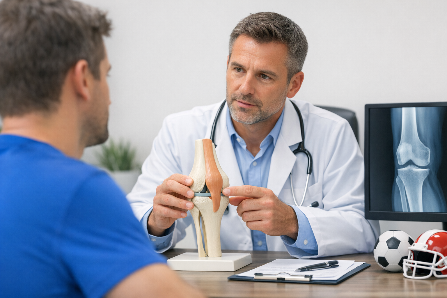
A radiology department already sits at a key decision point in knee injury care. MRI often answers questions about ligament morphology, edema, meniscal tears, cartilage injury, and bone bruising. However, many referrals also carry a second question: how unstable is the knee functionally? That is where objective knee laxity testing may add value.
In suspected ACL injury, post-reconstruction follow-up, or MRI-indeterminate instability, knee laxity testing can quantify anterior translation under controlled load. This gives referrers a structured measure rather than a purely descriptive impression. It is especially useful when the patient history suggests giving-way episodes, a pivoting mechanism, recurrent sport-related instability, or mismatch between MRI appearance and examination findings.
Radiology-led pathways are also increasingly multidisciplinary. A quantified instability result can sit alongside MRI findings, not in competition with them. That approach is particularly relevant in cases where MRI alone may not fully reflect functional instability, and in cases of rotational symptoms where rotational knee instability may be under-represented on static imaging.
Operationally, objective knee laxity testing works best when radiology defines three things clearly:
- Who should be referred for testing
- When the test should occur relative to MRI and clinic review
- How results will be reported back to the treating team
2. A step-by-step objective knee laxity testing workflow
The most effective model is a simple, referral-based pathway that fits existing MRI operations rather than disrupting them. A good knee ligament diagnostics workflow should be easy for referrers to understand and easy for radiology staff to execute.
2.1 Step 1 – Define referral indications
Objective knee laxity testing should be requested for focused clinical questions, not as a universal add-on for every knee MRI. Common indications may include:
- Suspected ACL injury with equivocal examination
- Borderline or partial ACL tear on MRI
- Pre-operative quantification of instability
- Post-operative follow-up after ACL reconstruction
- Persistent giving-way despite non-diagnostic imaging
- Research protocols requiring standardized dynamic knee instability assessment
For partial tears or borderline MRI findings, quantified testing may help clarify functional instability, but MRI remains complementary and is typically still needed to assess meniscus, cartilage, bone injury, and pre-operative planning when reconstruction is being considered. This is where a borderline ACL pathway can be especially useful.
2.2 Step 2 – Build timing into the imaging pathway
There are several workable models:
- Same-day model: MRI first, laxity testing after image acquisition if instability remains a referral question
- Triggered model: MRI report recommends objective knee laxity testing when findings are equivocal
- Combined sports knee clinic model: test performed before or after imaging within a coordinated service line
For most radiology practices, the triggered model is easiest to start. It keeps objective knee laxity testing focused on cases where the result may influence the next decision.
2.3 Step 3 – Standardize patient preparation
Preparation should be brief and reproducible. Staff should confirm:
- Injury side and symptom chronicity
- Prior ligament surgery
- Pain level and whether pain may limit relaxation
- Acute swelling or guarding
- Contraindications based on local protocol
Because muscle guarding can affect measurements, documentation of patient tolerance matters. In a radiology practice, this is not just a technical note. It helps clinicians interpret whether a low translation value reflects true stability or limited relaxation.
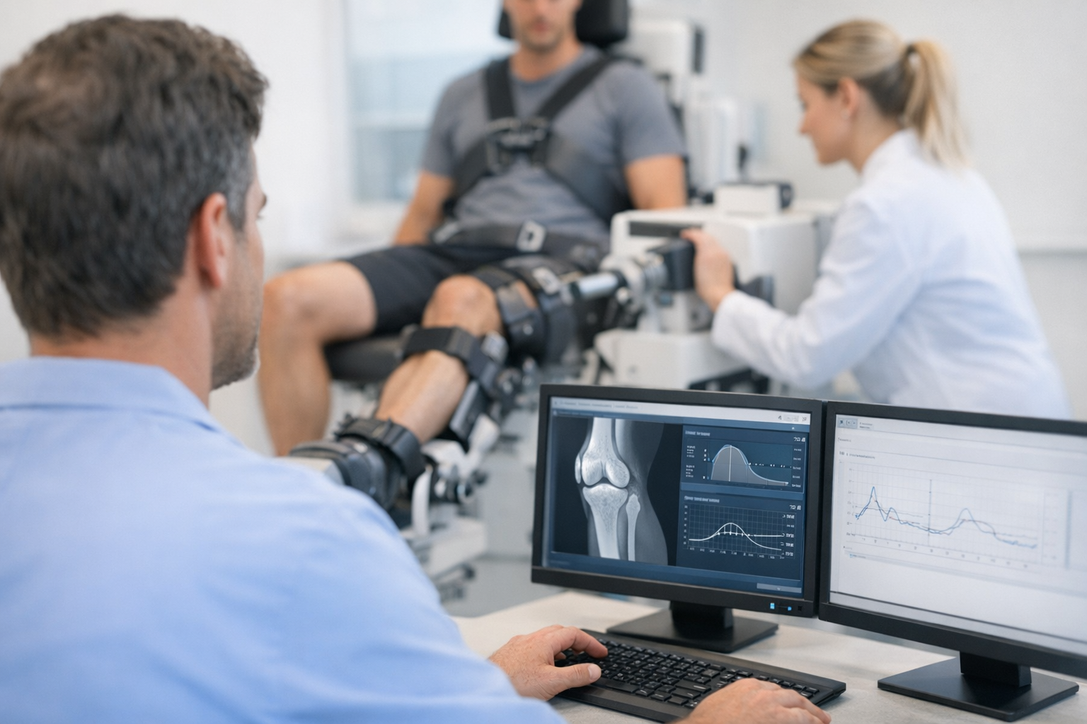
2.4 Step 4 – Measure both knees using a standard protocol
The core reporting metric for ACL-related testing is usually side-to-side anterior tibial translation. Bilateral comparison is often more informative than an isolated value from the symptomatic knee. A consistent protocol should specify patient position, fixation method, force progression, number of trials, and whether the practice reports maximal displacement, displacement at specific loads, or both.
This is where instrumented knee arthrometry becomes most useful: it reduces dependence on subjective examiner feel and creates a reproducible output for referring clinicians and researchers.
2.5 Step 5 – Integrate the result into the MRI report or addendum
The radiologist does not need to over-interpret the measurement. A concise functional summary is usually enough. For example:
- Measured side-to-side difference
- Whether the value suggests increased anterior laxity relative to the contralateral knee
- Whether interpretation is limited by pain, guarding, or poor tolerance
- Whether findings align or do not align with MRI appearance
Objective knee laxity testing is most valuable when it answers a practical clinical question. A report that simply lists a number without context is less useful than one that explains whether the quantified instability supports the referral concern.
2.6 Step 6 – Create a referral feedback loop
Radiology should review early cases with orthopaedic and sports medicine partners. This helps refine thresholds for use, reporting language, and triage. It also makes it easier to identify where objective knee laxity testing improves pathway efficiency, such as avoiding unnecessary repeat imaging in selected cases or fast-tracking review of clearly unstable knees.
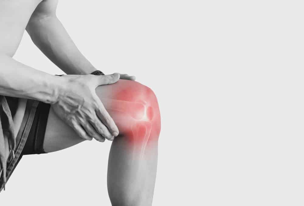
3. How to interpret results without overcalling instability
Interpretation needs discipline. Objective knee laxity testing can quantify instability, but it does not by itself diagnose every ligament pattern or define treatment. Results must be read with clinical examination, history, and MRI.
In ACL pathways, the most common question is whether the measured translation supports structural insufficiency seen on imaging. The answer is not always straightforward. Acute pain, hemarthrosis, limited relaxation, prior surgery, generalized laxity, and concomitant injury can all influence the reading.
Huang et al. (2025) examined associations between meniscal tear, tibial slope, static knee position, and anterior knee laxity in ACL-deficient patients. For a radiology workflow, the practical message is that measured laxity does not exist in isolation. Morphology and associated injuries may affect the instability profile, so objective knee laxity testing should be interpreted as one component of a broader assessment.
Moretti et al. (2025) explored anterior knee laxity and ACL MRI signals in healthy and reconstructed knees following exercise. That matters for scheduling and interpretation: knee state can change with activity, and radiology teams should keep testing conditions as standardized as possible when serial comparison is expected.
A short decision aid can help:
- If MRI clearly shows complete ACL rupture and the exam is concordant, objective knee laxity testing may be used mainly for baseline quantification or research.
- If MRI is borderline, partial, or clinically discordant, objective knee laxity testing may help clarify whether functional instability is present.
- If postoperative monitoring is the question, compare symptoms, graft appearance, and quantitative laxity over time rather than relying on one data point.
For broader interpretation frameworks, a quantified objective guide and an orthopaedic orthopaedics workflow can help teams align language across specialties.
4. Where instrumented and automated arthrometers fit
When a radiology practice decides to operationalize objective knee laxity testing, technology choice should be based on reproducibility, staff training burden, reporting output, and how easily the data can be integrated into the existing workflow. In this setting, an automated arthrometer radiology practice model may be attractive because it supports standardized load application and reduces operator variability compared with purely manual stress testing.
For example, GNRB arthrometer assessment and Dyneelax knee arthrometer platforms are designed for quantified anterior laxity evaluation and can support structured ACL laxity measurement in clinics that want reproducible side-to-side comparison. These systems should still be positioned as complementary to MRI and clinical examination, not as replacements.
In implementation planning, radiology leaders should ask:
- Can the device produce consistent bilateral measurements?
- Does the output support trend analysis and reporting?
- How much training is needed for operators to reach consistency?
- Can the service be scheduled in short appointment slots?
A practical starting point is to select one sports imaging pathway, one report template, and one trained operator group. Resources on the GNRB learning curve, comparative diagnostic tools, and robotic testing can help teams evaluate workflow fit.
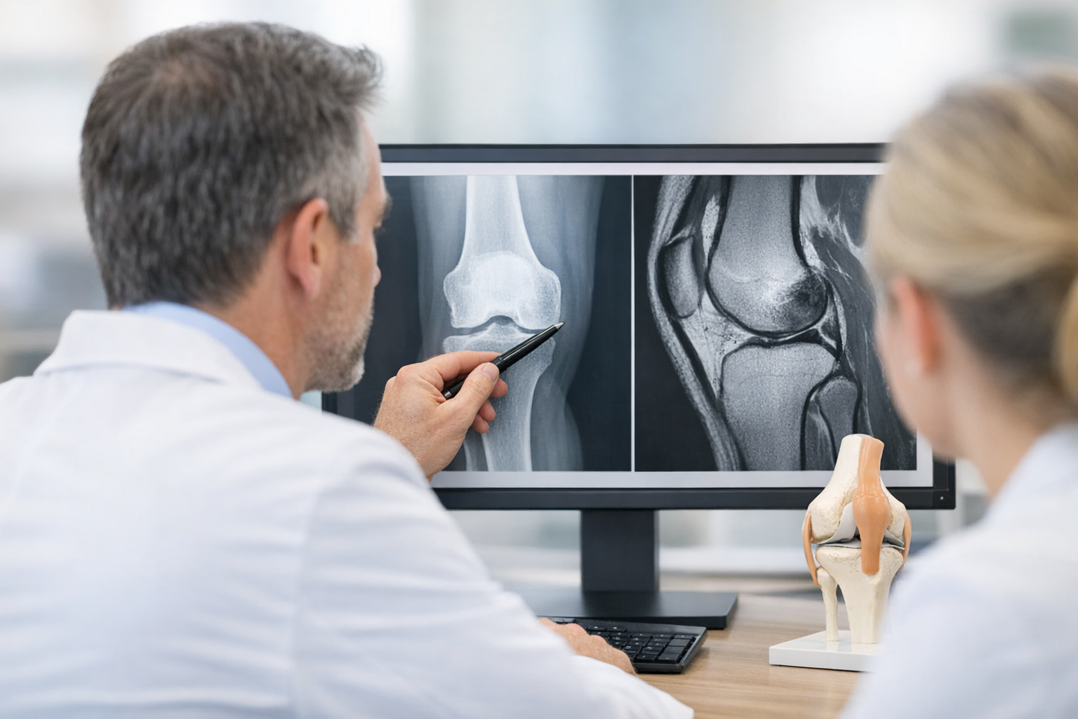
5. MRI integration, research opportunities, and common pitfalls
The strongest radiology pathway is one that integrates structural and functional data. MRI answers tissue-level questions. Objective knee laxity testing adds quantified function. Together, they may improve communication with surgeons, sports physicians, and rehabilitation teams.
Research is beginning to support this combined perspective. In reconstructed knees, Figueroa et al. (2025) reported a relationship between quantitative MRI UTE T2* of ACL autografts and BMI-normalized knee laxity within the first postoperative year. For radiology services, that type of GNRB laximeter MRI study context is important because it suggests that imaging biomarkers and objective laxity data may be meaningfully paired in follow-up protocols.
Similarly, Sinha et al. (2026) evaluated graft maturation and short-term clinical outcomes after ACL reconstruction techniques. While that trial is not a workflow paper, it reinforces a useful operational point: post-reconstruction assessment increasingly benefits from combining structural healing information with functional outcome measures, and objective knee laxity testing may contribute to that framework.
In combined ligament strategies, Chuang et al. (2025) reported short-term outcomes after ACL reconstruction plus anterolateral ligament reconstruction with suture tape augmentation. For radiologists, this is a reminder that persistent instability may have a rotational component, and MRI plus quantified testing may help frame when isolated anterior translation does not tell the whole story.
Common pitfalls in a radiology implementation include:
- Testing too early in painful, guarded acute knees
- Reporting numbers without context
- Using inconsistent bilateral technique
- Assuming anterior laxity fully captures pivoting symptoms
- Failing to define who owns follow-up interpretation
For MRI-linked pathways, radiology teams may also benefit from resources on an improving ACL tear detection strategy and a relevant MRI-laxity study context for reconstructed knees.
6. Key takeaways and next steps for implementation
A practical radiology pathway for objective knee laxity testing does not need to be complicated. It should be selective, standardized, and explicitly tied to clinical questions that MRI alone may not fully answer. In most practices, the best starting point is a targeted service for suspected ACL insufficiency, equivocal MRI findings, and postoperative follow-up.
The main principles are simple:
- Use objective knee laxity testing to complement MRI, not replace it
- Prioritize cases where quantified instability may change triage or clinical confidence
- Standardize bilateral technique and reporting language
- Interpret results with history, exam, and imaging
- Review early workflow performance with referrers and adjust
For radiologists and sports imaging teams, objective knee laxity testing can improve communication around ACL laxity measurement, support structured dynamic knee instability assessment, and make the overall knee ligament diagnostics workflow more actionable. The next step is usually operational, not theoretical: choose indications, train staff, pilot the protocol, and build a reporting template that makes objective knee laxity testing clinically useful from day one.
Clinical references (PubMed)
1) 2026 – Sinha et al. – ACL Reconstruction With Attachment-Sparing Hamstring Autograft Results in Earlier Graft Maturation and Better Short-Term Clinical Outcome in Comparison to Free Graft: A Randomized Controlled Trial – Am J Sports Med – DOI: 10.1177/03635465261455046 – PMID: 42310826 – PubMed
2) 2025 – Figueroa et al. – Relationship Between Quantitative MRI UTE T2* of ACL Autografts and BMI-Normalized Knee Laxity Within the First Year After ACL Reconstruction – Am J Sports Med – DOI: 10.1177/03635465251368393 – PMID: 40970673 – PubMed
3) 2025 – Huang et al. – The Association Between Concomitant Meniscal Tear, Tibial Slope, Static Knee Position, and Anterior Knee Laxity in ACL-Deficient Patients – Orthop J Sports Med – DOI: 10.1177/23259671251324186 – PMID: 40124192 – PubMed
4) 2025 – Moretti et al. – Anterior knee laxity and ACL magnetic resonance signals in healthy and ACL-reconstructed knees following exercise – Eur Rev Med Pharmacol Sci – DOI: 10.26355/eurrev_202507_37327 – PMID: 40748344 – PubMed
5) 2025 – Chuang et al. – Short-Term Outcome of Combined Anterior Cruciate Ligament Reconstruction and Anterolateral Ligament Reconstruction with Suture Tape Augmentation – J Clin Med – DOI: 10.3390/jcm14238283 – PMID: 41375585 – PubMed