A posterolateral corner injury is easy to under-recognize, especially when attention is pulled toward an ACL, PCL, or obvious fracture pattern. In practice, a posterolateral corner knee injury may present with subtle varus laxity, vague instability, or rotational symptoms that are attributed to pain inhibition or a more familiar ligament tear. That is why missed posterolateral corner injuries remain a recurring problem in acute trauma, sports medicine, and multiligament assessment. For orthopaedic surgeons, sports physicians, physiotherapists, and radiologists, the key is not just knowing the anatomy, but identifying the exam pattern, imaging clues, and decision points that reveal a clinically relevant posterolateral corner injury.
1. Why posterolateral corner injury is so often missed
The lateral side of the knee is anatomically complex and clinically noisy. A posterolateral corner injury rarely behaves like an isolated textbook lesion. More often, it appears alongside cruciate disruption, capsular injury, bony bruising, or swelling that limits a reliable examination.
The main reasons a posterolateral corner injury is missed include:
- Coexisting ligament trauma, especially in a triage algorithm setting where attention is focused on urgent exclusion of major instability patterns
- Pain and guarding, which may mask rotational and varus abnormalities early after injury
- Underuse of specific tests, particularly the dial test posterolateral corner assessment and comparison with the contralateral side
- MRI interpretation challenges, since edema, partial tearing, and slice orientation may obscure functional significance
- Failure to integrate exam findings into a structured objective knee examination
Clinically, the biggest trap is assuming that a confirmed cruciate tear explains everything. In many cases of multiligament knee injury PLC involvement, the cruciate lesion is only part of the instability problem. Super et al. (2026) highlights the complexity of multiligament knee injuries and reinforces why combined patterns require careful lateral and posterolateral assessment rather than isolated ligament thinking.
2. The anatomy that matters in posterolateral corner injury
To catch a posterolateral corner injury, clinicians need a practical rather than purely memorized view of the lateral side. The key stabilizers include the fibular collateral ligament, popliteus tendon, and popliteofibular ligament, with contributions from the capsule, biceps femoris complex, and iliotibial structures. In real terms, this is the system that resists varus stress, external rotation, and posterolateral rotatory instability knee patterns.
This is also where the distinction between isolated LCL and PLC injury and a broader posterolateral disruption becomes important. If only the LCL is considered, the clinician may underestimate rotational insufficiency. If only rotation is considered, an accompanying fibular-based lesion may be missed.
Radiologically, an anatomic framework can help. Saran et al. (2026) described the lateral knee as a “W” configuration, a concept that may improve recognition of how interconnected these structures appear on imaging. For radiologists and surgeons, that matters because a posterolateral corner injury is often better understood as a pattern, not a single torn structure.
For background on how different instability patterns present clinically, this overview of knee laxity can help frame why lateral and rotational findings should not be interpreted in isolation.
3. How to examine a suspected posterolateral corner injury
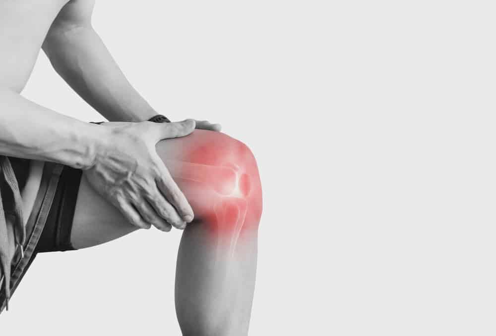
The bedside diagnosis of posterolateral corner injury depends on combining history, mechanism, alignment, and comparative testing. A patient may describe instability during cutting, deceleration, downhill walking, or pivoting rather than simple straight-line giving way. Varus thrust during gait, ecchymosis over the fibular head, and tenderness along the lateral joint line or popliteus region should raise suspicion.
3.1 Key clinical tests
Useful tests include:
- Varus stress testing at 0 and 30 degrees
- Dial test at 30 and 90 degrees
- Posterolateral drawer
- Reverse pivot shift
- External rotation recurvatum assessment
- Gait and standing alignment assessment
The dial test posterolateral corner remains especially valuable when interpreted carefully. Increased external rotation at 30 degrees may suggest isolated PLC involvement, while abnormal rotation at both 30 and 90 degrees can indicate combined PLC and PCL deficiency. This is not absolute, but it is a useful clinical pattern.
When rotational symptoms are prominent, clinicians should also consider how findings relate to broader instability pathways such as a high-grade pivot shift, since combined rotational laxity may reflect more than one injured stabilizer.
3.2 Common examination pitfalls
The exam is most vulnerable when done too early, too quickly, or without side-to-side comparison. A posterolateral corner injury may be underestimated if the patient is heavily guarded or if the examiner does not distinguish between pain-limited motion and true rotational opening.
Another common issue is assuming that a positive posterior drawer in a PCL-deficient knee explains all instability. In reality, Yang et al. (2026) explored rotational subluxation patterns on stress radiography in PCL-deficient knees, supporting the idea that rotational behavior may carry distinct clinical relevance beyond simple posterior translation.
For clinicians building a more quantitative assessment pathway, this summary of knee laxity testing can help organize how manual tests fit alongside objective measurements.
4. Imaging and objective testing in PLC knee injury diagnosis
PLC knee injury diagnosis should not rely on any single modality. Standard radiographs, long-leg alignment when appropriate, MRI, and stress imaging all contribute different information. MRI helps define injured structures and associated meniscal, cartilage, and osseous lesions, but functional instability may still require correlation with examination and stress-based assessment. This is particularly important in a posterolateral corner injury with borderline imaging findings or combined ligament injury.
Stress radiography can be practical in clinic when performed consistently. Lameire et al. (2026) described a simple stress radiograph technique for assessing laxity in multiligamentous knees, which is relevant for surgeons who need efficient ways to quantify asymmetry and support decision-making in a busy setting.
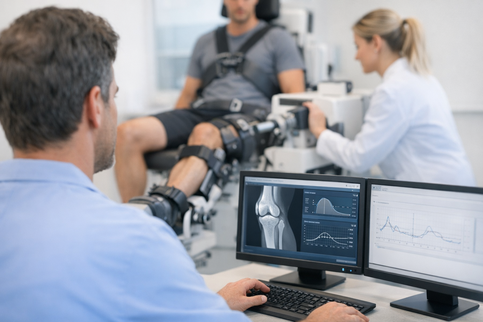
One sentence on technology integration: In complex rotational or multiligament assessment, a Dyneelax arthrometer may complement MRI and clinical examination by adding objective, side-to-side functional laxity data in selected cases of suspected posterolateral corner injury.
Where instrumented testing is used in practice, clinicians may also compare workflows involving a GNRB arthrometer or Dyneelax-based dynamic assessment depending on the suspected instability pattern and local protocol.
This matters because MRI remains complementary and often necessary for associated injuries and surgical planning, yet objective testing may help clarify functional instability when symptoms seem greater than static imaging suggests. That principle is discussed more broadly in MRI vs arthrometer pathways and in this review of the Dyneelax in diagnosis context.
For clinicians interested in reliability and reproducibility of instrumented assessment, this discussion of Dyneelax reliability is also relevant when building a repeatable follow-up process.
5. When posterolateral corner injury occurs with LCL, PCL, or multiligament trauma
A posterolateral corner injury becomes more consequential when it is part of a combined injury pattern. Persistent posterolateral laxity can compromise cruciate reconstruction outcomes, alter gait mechanics, and leave the patient with ongoing instability despite technically adequate surgery elsewhere.
Typical combined patterns include:
- LCL and PLC injury with varus and rotational instability
- PCL plus PLC deficiency with increased posterior and external rotation laxity
- ACL plus PLC injury with difficult-to-explain rotational symptoms
- Knee dislocation patterns where posterolateral corner injury is overshadowed by neurovascular priorities
In these settings, the diagnosis must move beyond naming torn structures and toward understanding the functional pattern. A posterolateral corner injury with associated cruciate damage may present as recurrent instability, graft overload risk, or unexplained asymmetry on return-to-sport testing.
Shahare et al. (2026) reported an unusual bilateral PLC presentation with asymmetric cruciate involvement, which is a useful reminder that clinically important posterolateral pathology does not always follow common expectations. Likewise, Study #3 reinforces that multiligament injuries should be approached systematically rather than by isolated lesion labels.
For clinics assessing complex instability patterns, multi-axis workflow thinking can be helpful, especially when a posterolateral corner injury may coexist with sagittal and rotational abnormalities.
6. A practical decision aid to reduce missed posterolateral corner injuries
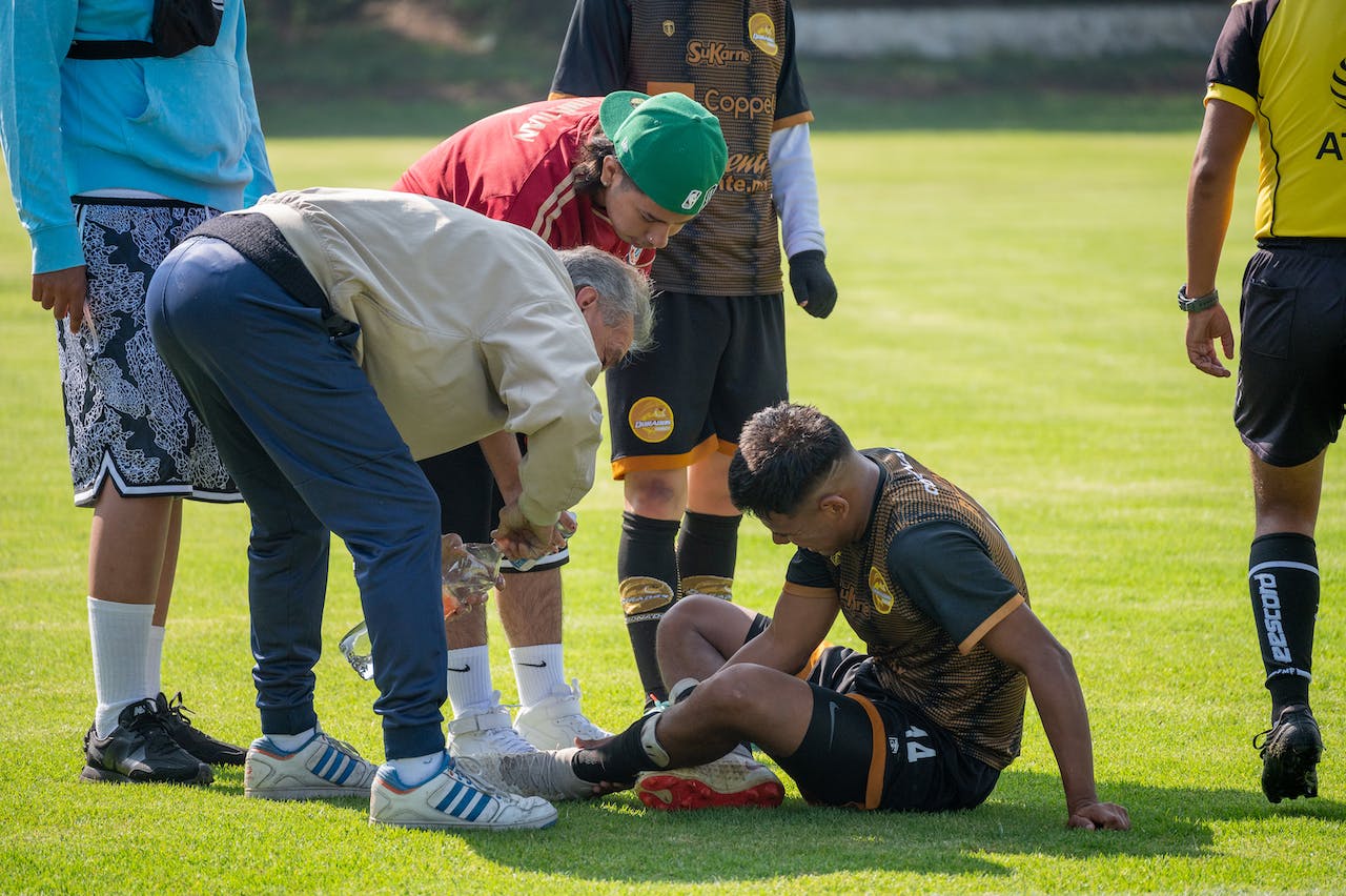
The simplest way to reduce missed posterolateral corner injuries is to standardize when you suspect them and what you do next. A posterolateral corner injury should move up the differential when the mechanism includes hyperextension, varus load, direct anteromedial blow, knee dislocation, or persistent rotational instability after cruciate injury.
Decision aid
- If lateral pain, varus opening, or rotational instability is present, actively screen for posterolateral corner injury rather than stopping at the first positive cruciate test.
- Compare both knees with varus stress and dial testing at 30 and 90 degrees.
- If findings are equivocal in the acute phase, repeat the exam after swelling and guarding improve.
- Use MRI to assess structural injury and associated pathology.
- Consider stress radiography or objective dynamic testing when function and imaging do not fully align.
- In any posterolateral corner injury associated with cruciate reconstruction planning, define whether residual posterolateral laxity is likely to affect treatment strategy.
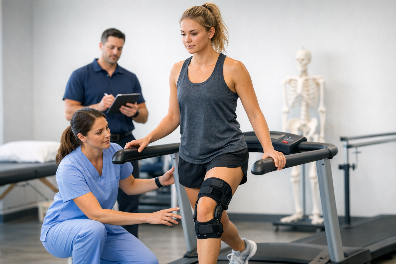
In rehabilitation and nonoperative follow-up, physiotherapists should also watch for recurrent giving way, avoidance of terminal extension, apprehension in pivoting tasks, and persistent asymmetry during functional testing. These can reflect unresolved posterolateral corner injury rather than deconditioning alone.
Broader context on dynamic testing is useful here, because functional instability may be most apparent when sagittal, varus, and rotational behavior are considered together.
7. Key takeaways and next steps
A posterolateral corner injury is commonly missed because it is complex, frequently combined with other ligament trauma, and easy to underestimate when examination is incomplete or imaging is interpreted without functional correlation. The most reliable safeguard is a structured approach: suspect it early, examine varus and rotational laxity carefully, compare both sides, and interpret MRI alongside the clinical picture.
For clinicians, the immediate next step is practical: build a repeatable assessment sequence for any knee with multiligament suspicion, unexplained rotational symptoms, or persistent instability after a known cruciate tear. A posterolateral corner injury should remain on the table until it is actively excluded, not passively overlooked.
Clinical references (PubMed)
1) 2026 – Lameire et al. – Assessment of Laxity in Multiligamentous Knee Injuries Using Stress Radiographs: A Simple Technique for a Busy Clinic. – Video J Sports Med – DOI: 10.1177/26350254251378514 – PMID: 41834844 – PubMed
2) 2026 – Yang et al. – Rotational Subluxation Patterns on Stress Radiography and Clinical Relevance in PCL-Deficient Knees: A 2D-3D Registration Study. – Orthop J Sports Med – DOI: 10.1177/23259671261445832 – PMID: 42293043 – PubMed
3) 2026 – Super et al. – Multiligament Knee Injuries. – J Bone Joint Surg Am – DOI: 10.2106/JBJS.26.00134 – PMID: 41984925 – PubMed
4) 2026 – Saran et al. – Crucial anatomic ensemble of the lateral knee: the ‘W’ configuration. – Clin Radiol – DOI: 10.1016/j.crad.2026.107288 – PMID: 41814107 – PubMed
5) 2026 – Shahare et al. – A Unique Presentation of Bilateral Posterolateral Corner Injury With Right Posterior Cruciate Ligament and Left Anterior Cruciate Ligament Tears. – Cureus – DOI: 10.7759/cureus.106046 – PMID: 42058347 – PubMed