In ACL tears hyperlax patients, the familiar heuristics behind Lachman grading, anterior drawer feel, and even pivot shift can break down because “normal” may already include large excursions. When generalized joint hypermobility or constitutional hyperextension exists, a large translation is not automatically pathologic, and a small side-to-side difference can still represent clinically relevant loss of restraint if both knees are highly lax. The clinical risk is twofold: overcalling instability (unnecessary surgery or extra procedures), or undercalling it (missed functional instability, delayed return-to-sport decisions, or graft choice and augmentation that does not match risk). This guide focuses on interpretation traps and on building better, patient-specific baselines that hold up across examiners and over time.
1. Why “high laxity” is not the same as “ACL failure”
Hyperlaxity is not a diagnosis, and it is not synonymous with ACL deficiency. It is a context that changes the pretest probability and the meaning of your exam findings. Before concluding that an exam reflects a torn graft or a new rupture in ACL tears hyperlax patients, clarify what “baseline” likely looked like.
1.1. Separate constitutional laxity from injury-driven instability
Start by documenting whether the patient has baseline knee laxity, recurvatum, or multi-joint features that raise the likelihood of constitutional looseness. A quick reference frame is the Beighton score and ACL discussion: Beighton is not knee-specific, but it helps you label the patient as “systemically lax” versus “isolated knee issue.”
For a concise refresher on terminology, it helps to align your team around what you mean by laxity versus instability, and then keep your notes consistent across visits.
1.2. Risk context: primary injury vs second injury is not identical
Risk signals from prospective cohorts are nuanced. For example, Fältström et al. (2025) reported that generalised joint hypermobility and excess knee hyperextension were associated with increased risk for a second ACL injury in female football players, but not for primary ACL injury. Clinically, that supports a more vigilant interpretation of “borderline” findings after return to sport in ACL tears hyperlax patients, especially when there is recurvatum and a history of reconstruction.
Similarly, in a prospective female athlete cohort, Pasanen et al. (2025) evaluated knee laxity and hypermobility among several candidate risk factors for non-contact ACL injury. Even when a study does not translate into a single clinic threshold, it reinforces a practical point: the more traits that increase laxity or loading vulnerability, the more you should rely on robust baselines and repeatable metrics rather than one-off “feel.”
1.3. Donor-site and construct choices can change measured laxity
Do not assume “more translation equals technical failure.” Graft configuration, harvest decisions, fixation, and rehabilitation all influence measured excursion. For example, Cristiani et al. (2025) reported comparable outcomes across hamstring tendon graft configurations, despite increased knee laxity with gracilis tendon harvesting. In ACL tears hyperlax patients, that kind of nuance matters when you interpret follow-up laxity in a patient who still reports confidence and has no pivot symptoms.
2. ACL tears hyperlax patients: interpretation pitfalls in manual tests
Manual tests remain central, but the signal-to-noise ratio is different when both knees move a lot. The goal is not to “downgrade” the exam, but to tighten the interpretation logic so you can defend your conclusion in ACL tears hyperlax patients.
2.1. Lachman: endpoint quality beats absolute excursion
In lax patients, the “amount” of translation is often less discriminative than endpoint quality, guarding, and symmetry under comparable relaxation. Revisit common interpretation errors using the Lachman test guide, especially the tendency to overcall a soft endpoint when hamstrings are active or the examiner is inadvertently adding rotation.
Practical pitfall: if you change tibial rotation between sides, you can create an artificial side-to-side difference. Standardize foot position and tibial rotation as much as possible before comparing.
2.2. Anterior drawer and translation feel: beware “false reassurance”
In chronic laxity, an anterior drawer can feel “not that bad” if the patient guards less on one side, if the meniscus provides a pseudo-block, or if the tibial slope and hamstring tone vary. For ACL tears hyperlax patients, interpret the drawer in context of symptoms (giving way, deceleration insecurity), functional tasks, and rotational signs.
2.3. Pivot shift: treat it as a rotational event, not a binary label
Pivot shift interpretation is particularly vulnerable to examiner technique, patient relaxation, and baseline laxity. A “trace” pivot in a very lax contralateral knee may be normal for that patient, while a “negative” pivot does not exclude symptomatic rotational instability if the patient is guarding or if you are not reproducing their provocation pattern.
When your exam and history diverge, revisit a bias-resistant process. Using a consistent framework for objective knee examination can reduce drift between clinicians and help you justify why you escalated testing or did not.
3. Better baselines: beyond contralateral comparison alone
Most clinicians anchor on the uninjured side. In ACL tears hyperlax patients, that is necessary but not sufficient because the contralateral knee may be unusually lax, previously injured, or developing compensations.
3.1. Use contralateral comparison, but qualify it
Contralateral knee comparison is still the fastest way to interpret a finding in real time, but document what might contaminate it: prior injury, apprehension, pain inhibition, or generalized hyperextension. When feasible, record the contralateral knee’s recurvatum and rotation because those features can “normalize” larger translations than you would accept in a stiffer athlete.
If your team uses classic arthrometer language, ensure everyone understands how thresholds were derived and what their limitations are in hyperlaxity. This overview of KT1000 side-to-side concepts is useful for aligning terminology, even if your clinic uses a different device.
3.2. Create a patient-specific baseline across time
A stronger baseline is a trajectory, not a single datapoint. In ACL tears hyperlax patients, repeat testing after swelling resolves and quadriceps control returns can change interpretation materially.
- Document the exam state: effusion grade, pain, guarding, and time from injury.
- Repeat key tests after prehab or early rehab when relaxation improves.
- Track function alongside stability. A resource on stability metrics over time can help reconcile “looks stable in the gym” versus “still gives way on the pitch.”
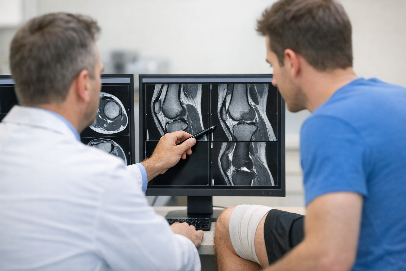
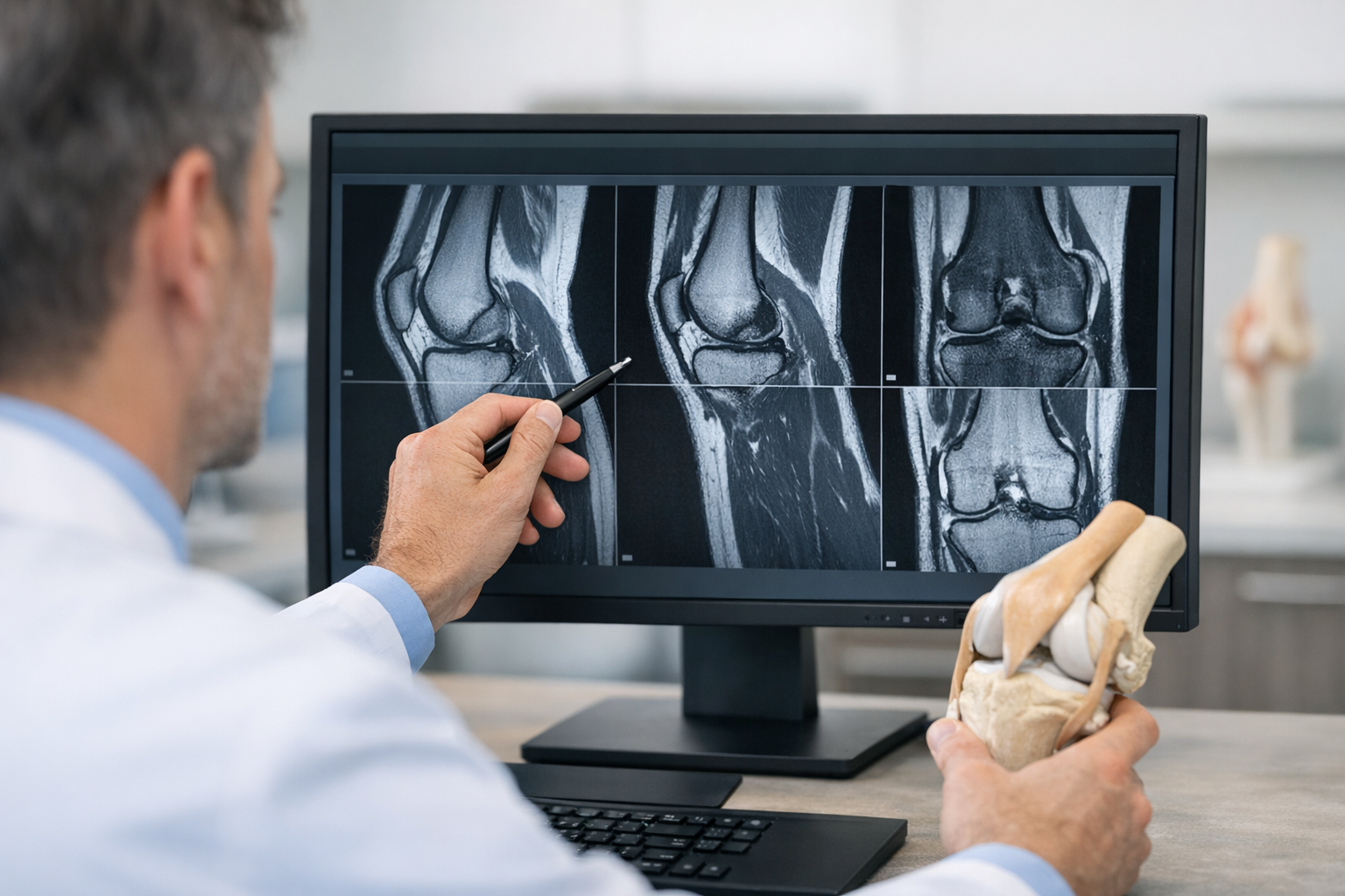
3.3. Clarify the MRI relationship in equivocal cases
In borderline presentations, the common failure mode is treating imaging as the only arbiter. MRI is essential for associated injuries and planning, but it does not always resolve functional instability, especially in partial tears or when fiber continuity looks preserved but restraint is compromised. In ACL tears hyperlax patients, an explicit workflow can reduce indecision: consider this workflow for integrating symptoms, exam, imaging, and instability confirmation.
When discussing test selection with referrers, keep the messaging balanced: quantitative instability measures can complement imaging. For a fuller comparison of roles, see MRI vs quantitative laxity testing (not as a replacement, but as an added functional layer).
4. Instrumented testing when baseline laxity is high
When manual tests are noisy, instrumented knee laxity testing can improve repeatability and help you move from “I think it is looser” to “the curve shifted.” This is especially useful in ACL tears hyperlax patients where both knees demonstrate large excursions and the diagnostic question becomes whether there is a meaningful asymmetry, altered mechanical response, or dynamic instability pattern.
4.1. Focus on measurement quality, not just millimeters
Two common errors are (1) treating a single force point as definitive and (2) ignoring curve shape. If available, incorporate an anterior tibial translation measurement across multiple loads, and document how the patient tolerated the test (guarding can artificially reduce translation). For clinics using robotic or instrumented systems, a standardized protocol can reduce operator dependence.
4.2. Stiffness and compliance can be more discriminative in hyperlaxity
In some hyperlax presentations, the clinically meaningful change is not the end-range translation but a change in compliance (how quickly translation increases with load). That is why stiffness-oriented interpretation is often emphasized for ACL tears hyperlax patients. A practical explanation of why stiffness and compliance metrics may outperform simple laxity thresholds can help with reporting and with surgical discussions.
4.3. Consider multi-axis capture when rotation drives symptoms
If the symptom is pivoting insecurity rather than straight-line giving way, pair anterior translation data with a rotational assessment plan. This multi-axis testing approach outlines how to think about combined planes when baseline laxity is high.
When your clinic uses dedicated devices, objective anterior translation and curve-based metrics can be captured with a GNRB arthrometer assessment or with a Dyneelax knee arthrometer as part of a clinician-led pathway that still incorporates history, exam, and MRI for associated injuries.
Postoperative interpretation also benefits from remembering that fixation choices do not necessarily change objective laxity at early follow-up. For example, Abram et al. (2026) reported no differences in objective knee laxity measurements or patient-reported outcomes between fixed and adjustable loop suspensory fixation at 1 year. In ACL tears hyperlax patients, that kind of evidence helps you avoid attributing every millimeter to a fixation choice rather than to biology, rehab state, or baseline phenotype.
5. Rotational instability: making pivot shift actionable in hyperlaxity
Rotational complaints are often what drives decision-making in ACL tears hyperlax patients, yet rotation is where baseline variability and examiner variability are highest. The solution is to structure what you are trying to learn: “Is there a clinically meaningful subluxation event that matches the patient’s complaint?”
5.1. A short decision aid for clinic
- Define the complaint: straight-line giving way, pivoting instability, or mistrust only.
- Standardize the exam: same tibial rotation, same relaxation strategy, same sequence.
- Anchor to baselines: contralateral knee, prehab-retest, and, if available, objective curves.
- Integrate MRI cautiously: confirm fiber status and, importantly, look for meniscus ramp/root tears, cartilage injury, bone bruising patterns, and other stabilizers.
- Escalate when mismatch persists: if symptoms suggest pivoting failure but the pivot is equivocal, consider structured dynamic testing and repeat exam under better relaxation.
5.2. What to do with a “big pivot” in a hyperlax athlete
A clearly positive pivot shift in a relaxed patient is still meaningful, even when the contralateral knee is loose. The harder scenario is when the pivot shift is “mild” but the athlete reports repeated episodes. In those cases, focus on modifiable contributors (neuromuscular control, fatigue exposure, landing mechanics) while you refine the stability baseline.
For surgical planning conversations, evidence about additional lateral procedures is evolving and is not specific to every hyperlax phenotype. In athletes with isolated ACL tears, El Abd et al. (2026) evaluated whether ACL reconstruction with lateral extra-articular tenodesis improves objective stability and functional outcomes. Use such trials to inform balanced counseling, while still individualizing to anatomy, sport demands, rotational findings, and tissue quality.
6. Closing: key takeaways and next steps
Key takeaways
- Hyperlaxity changes interpretation: focus on endpoint quality, symptom matching, and standardized technique rather than raw excursion alone.
- Do not let the contralateral knee become a false anchor. Qualify contralateral findings and build a serial, patient-specific baseline.
- Use MRI as a complementary tool for associated injuries and planning, and consider objective instability measures when function and imaging do not align.
- When rotation drives the complaint, make pivot shift assessment more reproducible by standardizing setup and integrating multi-plane thinking.
Next steps: If your current documentation cannot explain why you concluded “stable” or “unstable” in a lax phenotype, update your template to include baseline features (recurvatum, Beighton), contralateral qualifiers, and either serial or objective measurements so follow-ups remain comparable across clinicians.
7. Clinical references (PubMed)
1) 2025 – Fältström et al. – Generalised joint hypermobility and excess knee hyperextension are associated with an increased risk for second ACL injury, but not primary ACL injury, in female football players: A 5-year follow-up. – Knee Surgery, Sports Traumatology, Arthroscopy – DOI: 10.1002/ksa.70011 – PMID: 40923378 – PubMed
2) 2025 – Pasanen et al. – Knee laxity, joint hypermobility, femoral anteversion, hamstring extensibility and navicular drop as risk factors for non-contact anterior cruciate ligament injury in female athletes: A 4.5-year prospective cohort study. – Knee Surgery, Sports Traumatology, Arthroscopy – DOI: 10.1002/ksa.12625 – PMID: 39943896 – PubMed
3) 2026 – Abram et al. – No Differences in Objective Knee Laxity Measurements or Patient Reported Outcome Measures Between Fixed and Adjustable Loop Suspensory Fixation in Anterior Cruciate Ligament Reconstruction: 1-Year Results from the GAP Study, A Prospective, Double-Blinded, Randomized Trial. – J ISAKOS – DOI: 10.1016/j.jisako.2026.101123 – PMID: 42044694 – PubMed
4) 2026 – El Abd et al. – Does Anterior Cruciate Ligament Reconstruction with Lateral Extra-Articular Tenodesis Improve Objective Stability and Functional Outcomes in Athletes with Isolated Anterior Cruciate Ligament Tear? A Randomized Controlled Trial. – Journal of Knee Surgery – DOI: 10.1055/a-2778-8980 – PMID: 41544662 – PubMed
5) 2025 – Cristiani et al. – Comparable Outcomes Across 4 Hamstring Tendon Graft Configurations in ACL Reconstruction, Despite Increased Knee Laxity With Gracilis Tendon Harvesting. – Orthopaedic Journal of Sports Medicine – DOI: 10.1177/23259671251363585 – PMID: 40881380 – PubMed