Partial ACL injuries are common in pivoting sports, yet partial ACL tear diagnosis remains one of the most inconsistent decisions across orthopaedics, sports medicine, and radiology. The core problem is a mismatch between what patients feel (giving-way, rotational episodes, loss of trust) and what standard tests capture on the day, especially when only a portion of fibers is disrupted in a partial anterior cruciate ligament tear. MRI can depict fiber continuity, edema, and indirect signs, but functional instability can be subtle, load-dependent, and temporally variable. This comparison focuses on where MRI, clinical examination, and quantitative laxity tools agree, where they conflict, and how teams can reduce uncertainty without over-relying on any single modality.
1. Why partial ACL tears are missed in day-to-day practice
Missed or delayed recognition is rarely due to “bad care.” It more often reflects how partial injuries behave biologically and mechanically.
1.1 Anatomy and biomechanics: the injury is real, the signal is small
In partial tears, some bundle fibers may remain taut through parts of the range of motion, masking classic anterior translation. Acute pain, hemarthrosis variability, protective guarding, and reduced patient relaxation can all “normalize” tests that are highly sensitive in complete tears. This is why partial ACL tear diagnosis can look different at day 3 versus week 3, and why serial assessment matters.
1.2 Symptoms overlap with meniscus, MCL, and PLC patterns
Mechanical symptoms and episodic giving-way can arise from meniscal pathology, particularly posterior horn or ramp-type pathology, and from medial or posterolateral corner injuries. When clinicians anchor early on “meniscus” or “MCL sprain,” a partial ACL injury can be underweighted, even though the combined pattern is clinically important for return-to-sport decisions.
For a broader context on diagnostic pitfalls and false reassurance, see this overview.
1.3 The “borderline instability” trap
A patient can report high-demand instability while presenting with borderline translation on a single exam, a limited-quality MRI, or an MRI read that emphasizes “intact fibers.” In these cases, partial ACL tear diagnosis becomes a probability judgment, not a binary call, and the team benefits from explicitly documenting uncertainty and planning how to reduce it.
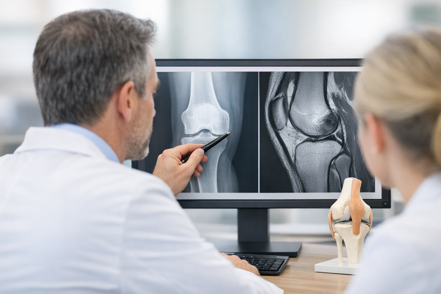
2. partial ACL tear diagnosis: comparing MRI, clinical tests, and functional instability
Clinically, the most useful comparison is not “MRI versus exam,” but “static structure versus dynamic function.” A high-quality pathway aligns these, rather than forcing one to overrule the other.
2.1 MRI: what it does well, and why partial tears are hard
MRI remains essential for evaluating associated injuries (meniscus, cartilage, bone bruising patterns) and for preoperative planning when reconstruction is considered. The challenge is that MRI accuracy for partial ACL tear depends on acquisition quality, reader experience, time from injury, and what the referrer asked the radiologist to specifically assess (bundles, tibial footprint, indirect signs).
Common reasons MRI undercalls partial disruption include:
- Volume averaging through oblique fibers and partial bundle continuity.
- Acute hemorrhage/edema obscuring fiber discontinuity.
- Chronic scarring that appears continuous but behaves functionally insufficient.
- Focus on “intact” fibers without correlating to laxity or rotational symptoms.
In equivocal imaging scenarios, structuring next steps helps. Many teams use a defined Workflow to reconcile symptoms, exam, and objective measures.
2.2 Clinical tests: fast, cheap, and still limited by context
Bedside testing remains indispensable, but partial injuries expose known constraints. The Lachman, anterior drawer, and pivot shift are influenced by relaxation, acute pain, examiner technique, and the presence of concomitant lesions. A useful checklist of exam components is summarized in tests, and deeper technique considerations are covered in this Lachman guide.
Key comparison points:
- Lachman: may show a soft endpoint or subtle increased translation, but can be “near-normal” in partial bundle preservation.
- Pivot shift: strongly relates to rotatory instability, but pivot shift test limitations include the need for relaxation, variable grading, and reduced sensitivity in acute guarding.
- Instrument-free grading: “+1 vs +2” is often too coarse for borderline cases, especially when return-to-sport decisions depend on small differences.
Because these findings are probabilistic, partial ACL tear diagnosis improves when clinicians document not only the grade, but also the testing conditions (pain, effusion, anesthesia or not, time since injury) and repeat the exam after swelling reduction or rehabilitation “unmasks” instability.
2.3 A practical comparison table for decision making
| Modality | Best at detecting | What it can miss in partial tears | When it is most helpful |
|---|---|---|---|
| MRI | Fiber morphology, edema, bone bruise patterns, meniscus and cartilage pathology | Functional insufficiency despite apparent continuity; subtle bundle disruption; “healed-looking” scar tissue | Suspected associated injuries; pre-op planning; confirming non-ACL pain sources |
| Clinical exam | Global knee status, endpoints, rotatory symptoms correlation | Guarding-related false negatives; coarse grading; inconsistency across examiners | Initial triage; follow-up trend; deciding what to test next |
| Quantitative laxity | Side-to-side instability quantification under standardized loads | May not localize the tear; can be confounded by generalized laxity or contralateral injury | Borderline imaging or exam; return-to-sport readiness discussions; longitudinal monitoring |
Used together, these tools reduce the “all or nothing” thinking that drives both over-treatment and missed instability. That integration is the real lever for better partial ACL tear diagnosis.
3. Rotational instability and PCL: the differentials that change interpretation
In borderline presentations, clinicians often focus only on anterior translation. Yet many “misses” occur because the dominant problem is rotational or multi-ligamentous.
3.1 Rotational instability: why it is under-captured
Patients frequently describe “twisting out” rather than pure forward shift. Rotatory laxity is influenced by lateral meniscus integrity, capsular structures, and the posterolateral corner. When a pivot shift cannot be elicited (pain, guarding), rotational instability may still be present. This is where a broader objective approach can help teams move beyond subjective grading; a useful examination framework can standardize what is recorded and reduce inter-clinician variability.
3.2 PCL and multi-ligament patterns: don’t let one sign “explain everything”
A subtle posterior sag, a positive quadriceps active test, or altered tibial station can distort perceived anterior translation and confuse partial ACL tear diagnosis. In addition, MCL injury can both mask and amplify perceived laxity depending on testing position, while PLC deficiency can present as rotational giving-way with less impressive anterior translation.
When the story suggests instability but classic ACL tests are borderline, explicitly re-check:
- Effusion and pain inhibition
- Tibial station (posterior sag)
- Varus and dial test asymmetry (PLC screen)
- Medial opening at 30 degrees (MCL)
- Meniscal mechanical signs and joint line tenderness
Conceptually, this is also why a purely “static image” approach can miss functionally important laxity patterns. See the discussion on dynamic assessment concepts when symptoms and imaging diverge.
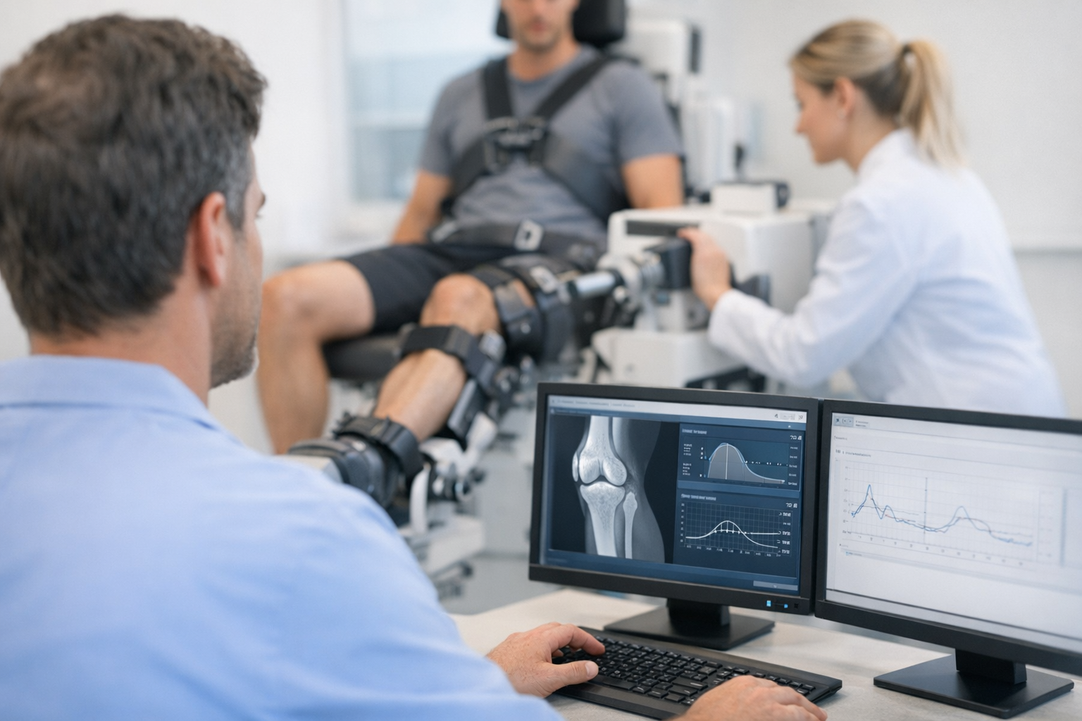
4. Objective laxity testing in MRI-equivocal knees
This is the step that often reduces “diagnostic stalemates.” Objective tools do not replace MRI, but they can add functional information by quantifying instability under standardized conditions.
4.1 What to measure: translation, differential, and load response
For clinical decision-making, the most actionable metrics typically include:
- side-to-side differential laxity (injured vs contralateral knee) rather than an isolated absolute number
- dynamic anterior tibial translation behavior across increasing loads (not only a single endpoint)
- Quality of the endpoint and repeatability across trials
In practice, these principles align with broader guidance on laxity workflows. The goal is not a single “magic cutoff,” but to reduce uncertainty and improve the confidence of partial ACL tear diagnosis in the specific patient in front of you.
4.2 Evidence snapshot: arthroscopic validation and clinical realism
A key comparative paper is Cojean et al. (2023), a prospective diagnostic study with arthroscopic validation that evaluated a GNRB laximeter alongside MRI in routine practice for complete and partial ACL tears (214 patients). Clinically, this is often summarized as GNRB vs MRI Cojean 2023, and it is particularly relevant when MRI wording is “partial tear suspected” yet the functional question remains unanswered.
Importantly, this is complementary positioning: MRI is still typically needed to assess meniscus and cartilage, while objective laxity metrics may help clarify borderline or discordant cases of partial ACL tear diagnosis.
4.3 Implementation in clinic: reliability, learning curve, and reporting
When teams add objective knee laxity measurement, they should define who performs the test, how knee position is standardized, and how results are integrated into reports. Training matters; practical considerations are discussed in learning resources.
Where available, instrumented arthrometer testing may be performed with devices such as the GNRB arthrometer or the Dyneelax arthrometer, particularly when clinical examination and imaging do not fully explain the patient’s instability.
For a device-and-imaging comparison that is directly relevant to equivocal cases, see MRI-vs.
4.4 A short decision aid for reducing uncertainty
- Start with probability: document suspected partial tear vs functional instability vs alternative diagnosis.
- Reconcile contradictions: if symptoms suggest giving-way but MRI is equivocal, do a standardized re-exam after effusion and pain improve.
- Quantify: use side-to-side and load-response metrics to support or weaken the ACL hypothesis.
- Search for “instability multipliers”: meniscal ramp/roots, MCL, PLC, generalized laxity, contralateral injury.
- Decide the next node: rehab with close monitoring, repeat imaging, or surgical consultation depending on instability, sport demands, and associated injuries.
In urgent or resource-limited settings, a standardized triage pathway can help ensure borderline cases do not fall through follow-up.
5. Key takeaways and next steps for clinicians
Reducing missed partial injuries is mainly about upgrading how uncertainty is handled. The best-performing teams treat partial ACL tear diagnosis as an iterative synthesis of symptoms, exam quality, imaging, and function.
- MRI is necessary but not sufficient in many partial or borderline cases: it is excellent for associated injuries and planning, but less direct for function.
- Clinical tests remain central, yet grading variability and guarding mean “negative” does not always mean “stable,” especially for rotational events.
- Quantifying instability (side-to-side and load response) can narrow the gray zone and make multidisciplinary discussions more precise.
Three evidence-informed cautions that influence “next steps” decisions:
- Nonoperative pathways can be appropriate in select patients, but outcomes and progression risk are context-specific; see Hannon et al. (2026) for pediatric nonoperative management outcomes in partial ACL tears.
- MRI can miss clinically important concomitant pathology; for example, meniscal injury patterns and ramp-associated lesions are a recurring theme in imaging misses, highlighted by Moran et al. (2026).
- Getting the initial decision wrong can carry long-term consequences in high-demand athletes and revision scenarios; relevant outcomes perspectives are discussed in Rubin et al. (2026) and in the MOON cohort findings from Magnussen et al. (2026).
Next steps in practice:
- If MRI is equivocal, document “suspected partial tear vs functional instability” explicitly and plan what will reduce uncertainty at the next visit.
- Reassess after effusion decreases, and consider adding quantified metrics when translation is borderline or symptoms are mainly rotational.
- Communicate clearly with radiology about clinical suspicion and suspected associated injuries (meniscus, PLC, MCL, cartilage).
6. Clinical references (PubMed)
1) 2023 – Cojean et al. – GNRB® laximeter with magnetic resonance imaging in clinical practice for complete and partial anterior cruciate ligament tears detection: A prospective diagnostic study with arthroscopic validation on 214 patients. – Knee – DOI: 10.1016/j.knee.2023.03.017 – PMID: 37172464 – PubMed
2) 2026 – Hannon et al. – Outcomes of Initial Nonoperative Management of Partial Anterior Cruciate Ligament Tears in Pediatric Patient. – J Pediatr Soc North Am – DOI: 10.1016/j.jposna.2026.100330 – PMID: 41908101 – PubMed
3) 2026 – Moran et al. – Concomitant Lateral Meniscal Tears in Pediatric and Adolescent Patients Undergoing Combined Medial Meniscal Ramp Lesion Repair and Anterior Cruciate Ligament Reconstruction Are Frequently Missed on MRI, Are Often Vertical or Root Tears, and Are Usually Repaired: A Multicenter Study. – Orthop J Sports Med – DOI: 10.1177/23259671251396140 – PMID: 41732223 – PubMed
4) 2026 – Rubin et al. – The impact of isolated ACL and MCL injuries on career longevity and performance metrics in elite rugby union players. – J Orthop – DOI: 10.1016/j.jor.2026.02.051 – PMID: 41736904 – PubMed
5) 2026 – Magnussen et al. – Having a Revision ACL Reconstruction Is Worse Than Tearing the Contralateral ACL and Undergoing Reconstruction: A MOON Cohort Study. – Am J Sports Med – DOI: 10.1177/03635465261416923 – PMID: 41696862 – PubMed