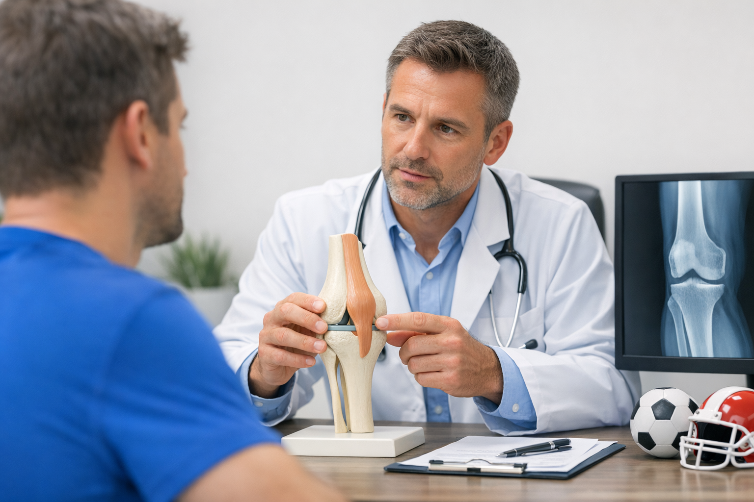
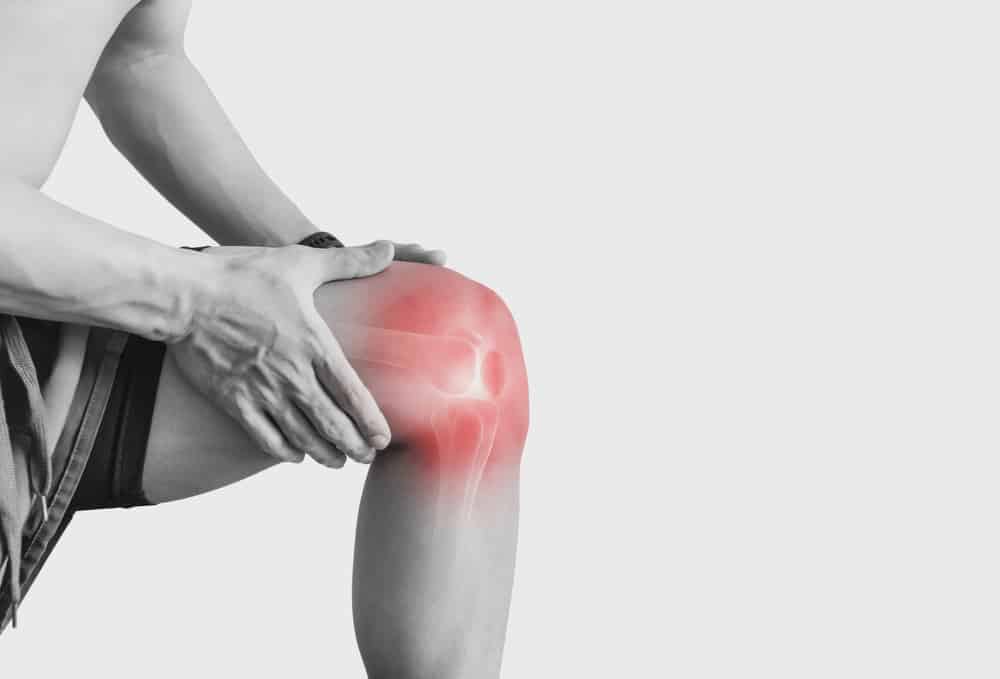
After surgery, a stable-feeling knee does not always equal a stable knee, and a “loose” measurement does not always mean a failing graft. ACL reconstruction follow-up testing helps clinicians distinguish normal recovery variation from clinically relevant instability by combining symptoms, examination, functional progress, and selected imaging. This matters when you are counseling athletes who feel uncertain, when objective findings lag behind rehabilitation, or when knee laxity after ACL reconstruction raises concern about graft behavior. The goal is not to chase a single number, but to make decisions that are timed, reproducible, and clinically responsible, including when to intensify rehab, pause progression, investigate contributing anatomy, or initiate a failure workup.
1. ACL reconstruction follow-up testing workflow: who, when, and what it answers
Use ACL reconstruction follow-up testing as a decision workflow rather than a one-off “check.” In practice, each follow-up visit should answer one primary question, then select the minimum set of tests needed to support the decision.
Define the decision before choosing tests:
- Reassure and progress (normal trajectory, readiness for higher loads)
- Explain mismatch (good strength but instability symptoms, or vice versa)
- Triage imaging (new trauma, mechanical symptoms, effusion, persistent giving-way)
- Identify risk (recurrent instability, contralateral issues, high-demand sport, abnormal kinematics)
It helps to document each timepoint as an “objective exam snapshot.” A structured template based on objective knee examination in orthopaedics supports longitudinal comparison, reduces narrative drift between providers, and makes subtle change easier to detect.
Two reminders keep ACL reconstruction follow-up testing clinically grounded:
- Prioritize symptoms and function when the exam is borderline.
- Prioritize objective stability when symptoms are vague, fear-driven, or heavily load-dependent.
2. Step 1: Timepoint-based planning (knee stability testing timeline after ACL surgery)
A practical knee stability testing timeline after ACL surgery aligns what you measure with what tissue biology and motor control can realistically support. ACL reconstruction follow-up testing is most useful when repeated at consistent milestones using consistent methods.
2.1 Early phase (approximately 0 to 12 weeks): establish a baseline and screen red flags
Early ACL reconstruction follow-up testing is less about “final stability” and more about building a baseline for later comparison:
- Effusion, pain, extension deficit, quadriceps inhibition, gait deviation
- Wound status and early complications
- Gentle anterior drawer or Lachman quality (avoid over-interpreting small changes)
Pitfall: early guarding and arthrogenic muscle inhibition can distort exam quality and patient-reported instability. Consider the behavioral overlay described in AMI and kinesiophobia when the story (fear, avoidance) does not match the physical findings.
2.2 Mid phase (approximately 3 to 9 months): quantify stability alongside strength and control
This is when most clinical decisions intensify: running initiation, cutting progressions, and the first serious “return” conversations. Repeatable ACL reconstruction follow-up testing here can reveal whether change is improving, plateauing, or drifting in the wrong direction.
For allograft patients, structural changes can be dynamic across time. Serial morphology is one reason to keep assessment consistent rather than ad hoc, as highlighted by You et al. (2026) (serial graft diameter changes following allograft ACL reconstruction).
2.3 Late phase (9 months to 5 years): detect drift, compensations, and contralateral patterns
Late ACL reconstruction follow-up testing often shifts from “is the graft intact?” to “is the knee functioning stably in sport?” Long-term follow-up can reveal that passive laxity and functional stability do not always align, consistent with Beveridge et al. (2026), which discusses passive laxity with functional stability and potential bilateral dynamic compensatory mechanisms at long-term follow-up.
At multi-year timepoints, consider structural pathways (tunnel changes, maturation) and function together. For example, Tang et al. (2026) links mid-term tibial tunnel enlargement and graft maturation dynamics to knee function over 5 years, reinforcing that “structure” and “performance” should be interpreted together.
3. Step 2: Build the follow-up test battery (laxity, pivot shift, and function)
Use three complementary domains in ACL reconstruction follow-up testing:
- Passive/anterior stability (how the tibia translates under standardized load)
- Rotational instability (pivot shift behavior and control-dependent giving-way)
- Functional capacity (strength, hop tasks, landing mechanics, endurance)
Start by standardizing the basics from your chosen methods: document technique, knee flexion angle, stabilization, patient relaxation, and whether the endpoint is firm or soft.
3.1 Passive laxity: focus on symmetry and change, not a single absolute value
In routine follow-up, the clinically useful unit is often the side-to-side difference anterior tibial translation, interpreted in context of symptoms, endpoint quality, and trajectory over time. This is where “trend” matters: repeated ACL reconstruction follow-up testing is typically more informative than an isolated measurement.
Interpretation tips (contextual, not absolute thresholds):
- Stable trend with good function can support progression even if some asymmetry persists.
- Worsening trend across visits, especially after a giving-way event, raises concern for graft stretch, fixation issues, or new injury.
- Unexpected improvement may reflect reduced guarding and better relaxation rather than a true mechanical change.
3.2 Rotational instability: do not skip the pivot shift conversation
Pivot shift assessment after ACL reconstruction remains central because many “instability” complaints are rotational and activity-dependent rather than purely anterior. Your follow-up documentation should capture:
- Pivot shift grade (and whether it is under anesthesia vs awake)
- Apprehension/guarding and hamstring dominance patterns
- Dynamic valgus, trunk control, and cutting mechanics
Graft type and neuromuscular strategy may influence dynamic movement even when gross kinematics appear similar. Vendrig et al. (2026) reports similar dynamic tibiofemoral movements during jump-landing and walking but distinct hamstrings strength across ACL reconstruction autograft types at mid-term follow-up, supporting the idea that a “normal-looking” task can still hide clinically meaningful capacity differences.
3.3 Functional capacity: pair stability with readiness
Return decisions should not be made from laxity alone. Incorporate functional performance tests after ACL reconstruction such as hop test clusters, isokinetic or dynamometry strength (quadriceps and hamstrings), and task-quality assessment (single-leg squat, landing, deceleration). When the clinical question is sport clearance, make sure your pathway explicitly includes return to sport testing after ACL surgery and not just time since surgery.
To prevent “rehab milestone drift,” compare performance to stability metrics and symptom behavior using milestones as a cross-check, particularly when the athlete feels ready but objective measures lag.
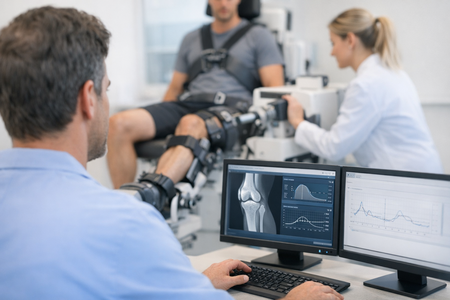
4. Objective laxity and rotational measurement in follow-up (complements MRI, not a replacement)
One role of ACL reconstruction follow-up testing is to quantify what hands-on grading can only estimate, particularly when the exam is limited by patient guarding, body habitus, or variability between examiners. In selected pathways, objective dynamic laxity testing can add functional information about how the knee responds to load and how symmetry evolves over time.
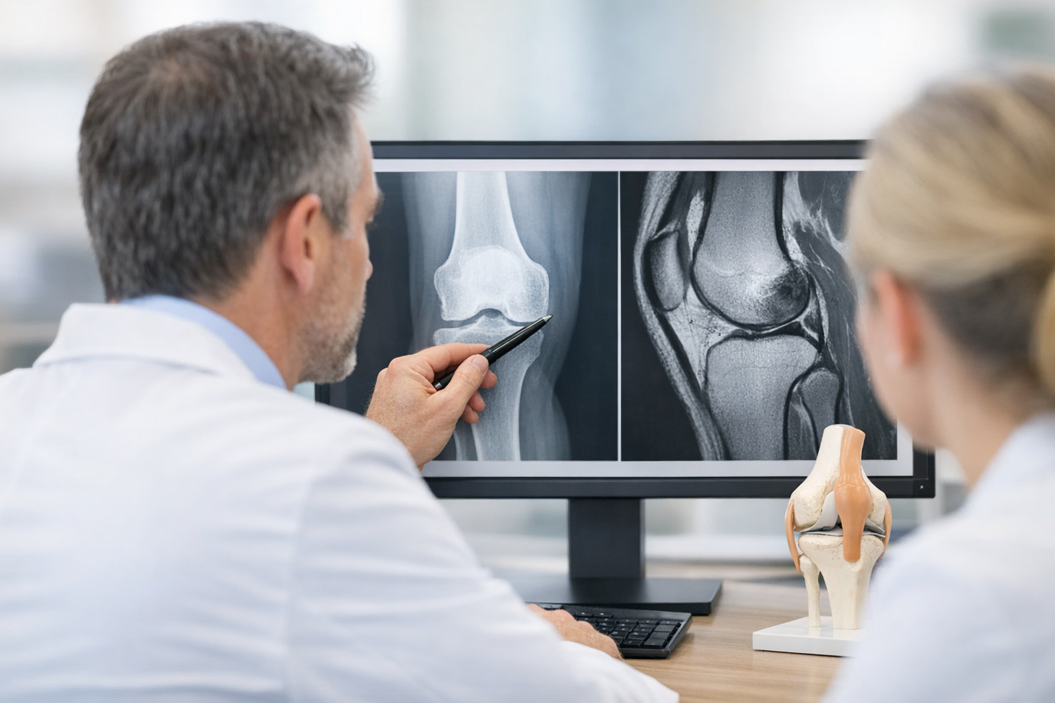
Positioning matters: instrumented testing should be framed as complementary to clinical examination and MRI, not a substitute. MRI remains important for assessing meniscus, cartilage, bone bruising, tunnel position issues, cyclops lesions, and other contributors to symptoms and for surgical planning when indicated.
Clinics commonly integrate instrumented anterior translation measurement and, when available, multi-axis assessment to better reflect functional instability. A multi-direction approach is outlined in multi-axis follow-up pathways.
When instrumented testing is used, choose metrics that are reproducible and clinically interpretable. Some teams track stiffness and compliance characteristics rather than translation alone because they may better reflect graft behavior and patient relaxation; see the rationale behind compliance and stiffness approach.
Examples of instrumented systems used in clinical follow-up include the GNRB arthrometer and the Dyneelax knee arthrometer, which can support repeatable documentation of anterior tibial translation and side-to-side response under standardized loading.
If imaging is part of the workup, ensure measurement reliability for bony morphology and kinematics if using advanced modalities. For example, VanTienderen et al. (2026) evaluates inter- and intra-observer reliability of measuring posterior tibial slope, anterior tibial translation, and tibiofemoral rotation using weight bearing CT, underscoring the value of standardized imaging measurement methods when anatomy is driving decisions.
5. Step 3: Interpret change, monitor graft integrity, and decide the next clinical action
At this stage, ACL reconstruction follow-up testing is about interpretation: what does the pattern mean, and what should change in management today? Use a simple three-lane interpretation model: expected recovery, needs targeted rehab modification, or needs further investigation.
5.1 Decision aid: what to do with a new or worsening side-to-side gap
When the side-to-side difference anterior tibial translation increases compared with prior visits, use a stepwise check before labeling it as failure:
- Confirm test validity: same technique, patient relaxed, no effusion spike, comparable knee angle, no acute pain limitation.
- Re-check the story: any new pivoting injury, giving-way, “pop,” swelling, or loss of trust?
- Pair with rotation: repeat pivot shift and assess whether rotational symptoms match translation change.
- Assess function: strength symmetry, quality of deceleration/landing, fatigue effects, and psychological readiness.
- Escalate appropriately: consider MRI (and other imaging if anatomy is suspected), and consider referral for surgical opinion if findings suggest structural compromise.
This is the clinical zone where graft integrity monitoring after ACL reconstruction should be explicit in the record: trend direction, endpoint feel, rotational findings, and whether the patient reports instability during sport-specific tasks.
For a structured way to interpret post-op laxity change as benign stretch vs clinically meaningful failure, use the stretch interpretation workflow.
5.2 When symptoms and laxity do not match
Mismatches are common in ACL reconstruction follow-up testing:
- Loose but stable function: may reflect adaptation, bilateral strategies, or measurement context, consistent with the long-term compensation discussion in Beveridge et al. (2026).
- Tight measurements but instability complaints: consider rotational instability drivers, meniscal deficiency, chondral pain, AMI, fear avoidance, or poor movement control.
If the complaint is rotational giving-way with a low-grade translation exam, revisit pivot shift assessment after ACL reconstruction (including guarding) and consider multi-axis testing and/or imaging to evaluate secondary stabilizers and associated pathology.
5.3 Integrate imaging without over-calling graft problems
Imaging can clarify structure, but clinical context remains key. MRI is useful for associated injuries and graft appearance, while objective stability measures can provide a functional counterpart. Early post-op relationships between graft signal and measured laxity have been explored in quantitative MRI graft signal and laxity, which is relevant when the clinical question is whether a “concerning-looking” graft signal aligns with functional stability.
In longer follow-up, structural and tunnel changes may coexist with acceptable function. The 5-year morphological dynamics described by Tang et al. (2026) are a reminder to avoid one-dimensional conclusions based on a single modality.
5.4 Return-to-sport clearance: tie stability to performance criteria
For sport clearance, ACL reconstruction follow-up testing should support a clear yes, no, or not-yet decision. A practical structure:
- Stability: translation symmetry trend, endpoint quality, and rotational control (pivot shift and task-based symptoms).
- Capacity: quadriceps and hamstrings strength, rate-of-force development where available, and hop task cluster results.
- Quality: landing mechanics, fatigue tolerance, and sport-specific deceleration and change-of-direction control.
- Context: sport, position, exposure, and athlete risk tolerance.
Use objective criteria to prevent premature clearance, especially when time-based expectations dominate. See stability-linked return decisions and benchmarks that align objective stability with return-to-sport frameworks.
5.5 When findings suggest failure or a revision pathway
Escalate from routine ACL reconstruction follow-up testing to a failure evaluation when you have a compatible history (new pivot injury or repeated giving-way), worsening objective instability across visits, or persistent high-grade rotational instability despite adequate rehab adherence. In these cases, consider a structured revision workup pathway that incorporates imaging, alignment and slope assessment, meniscal status, and activity demands.
6. Key takeaways and next steps
ACL reconstruction follow-up testing is most valuable when it is repeatable, timepoint-appropriate, and explicitly linked to a clinical decision. A minimal, clinic-ready approach is to track (1) symptoms and swelling, (2) anterior and rotational stability, and (3) functional capacity and movement quality, then interpret the trend rather than a single visit result.
- Use trends: repeated measures reduce noise and clarify whether change is expected recovery, plateau, or deterioration.
- Match the test to the question: pivot symptoms require rotational assessment, not translation alone.
- Integrate, do not substitute: objective stability measures complement clinical exam and MRI when the pathway requires deeper clarification.
Next steps for clinicians: document a standardized battery, decide which milestones trigger repeat stability measures, and predefine what change triggers imaging, rehab modification, or referral. Next steps for patients: report any new giving-way events, swelling spikes, or confidence collapse during pivoting tasks, as these change the meaning of follow-up findings.
Clinical references (PubMed)
1) 2026 – Tang et al. – Mid-Term Tibial Tunnel Enlargement and Graft Maturation After ACL Reconstruction: A 5-Year Follow-Up Linking Morphological Dynamics to Knee Function. – Orthopaedic Surgery – DOI: 10.1111/os.70287 – PMID: 41854376 – PubMed
2) 2026 – Beveridge et al. – Passive Laxity With Functional Stability Reveals Potential Bilateral Dynamic Compensatory Mechanisms in Patients Undergoing ACL Reconstruction at Long-term Follow-up. – Orthopaedic Journal of Sports Medicine – DOI: 10.1177/23259671251414857 – PMID: 42004453 – PubMed
3) 2026 – You et al. – Serial Changes in Graft Diameter Following Anterior Cruciate Ligament Reconstruction Using Allograft. – Cartilage – DOI: 10.1177/19476035261441851 – PMID: 42011883 – PubMed
4) 2026 – Vendrig et al. – Similar dynamic tibiofemoral movements during jump-landing and walking but distinct hamstrings strength across ACL reconstruction autograft types at mid-term follow-up. – Knee Surgery, Sports Traumatology, Arthroscopy – DOI: 10.1002/ksa.70275 – PMID: 41562143 – PubMed
5) 2026 – VanTienderen et al. – Inter- and intra-observer reliability of measuring posterior tibial slope, anterior tibial translation, and tibiofemoral rotation utilizing weight bearing CT. – European Journal of Radiology – DOI: 10.1016/j.ejrad.2026.112877 – PMID: 42013582 – PubMed