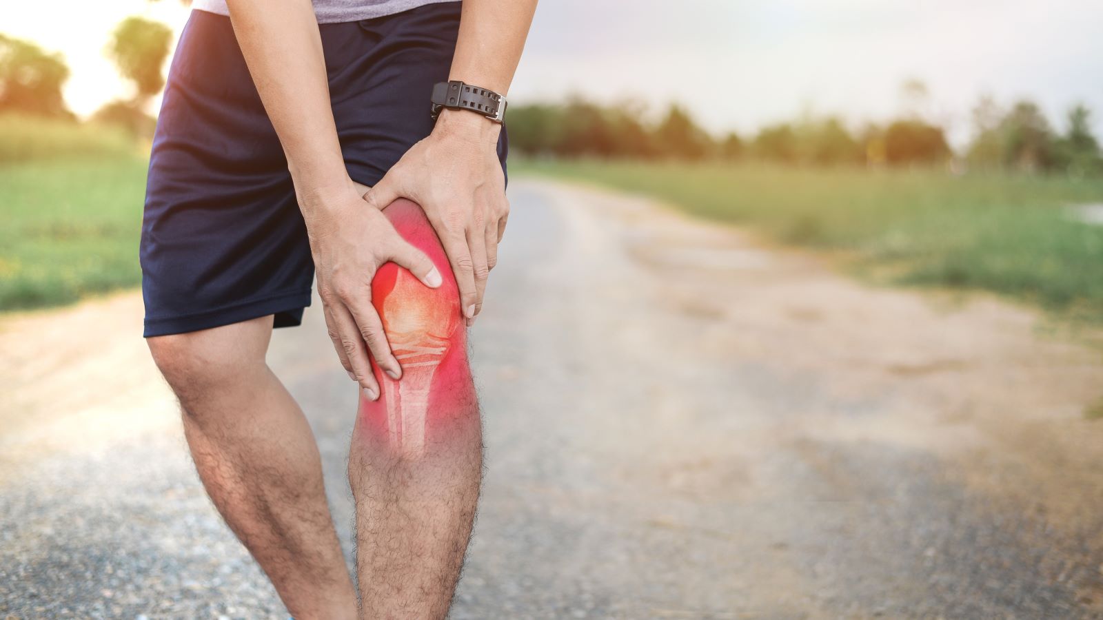
Introduction to the KT1000 Arthrometer
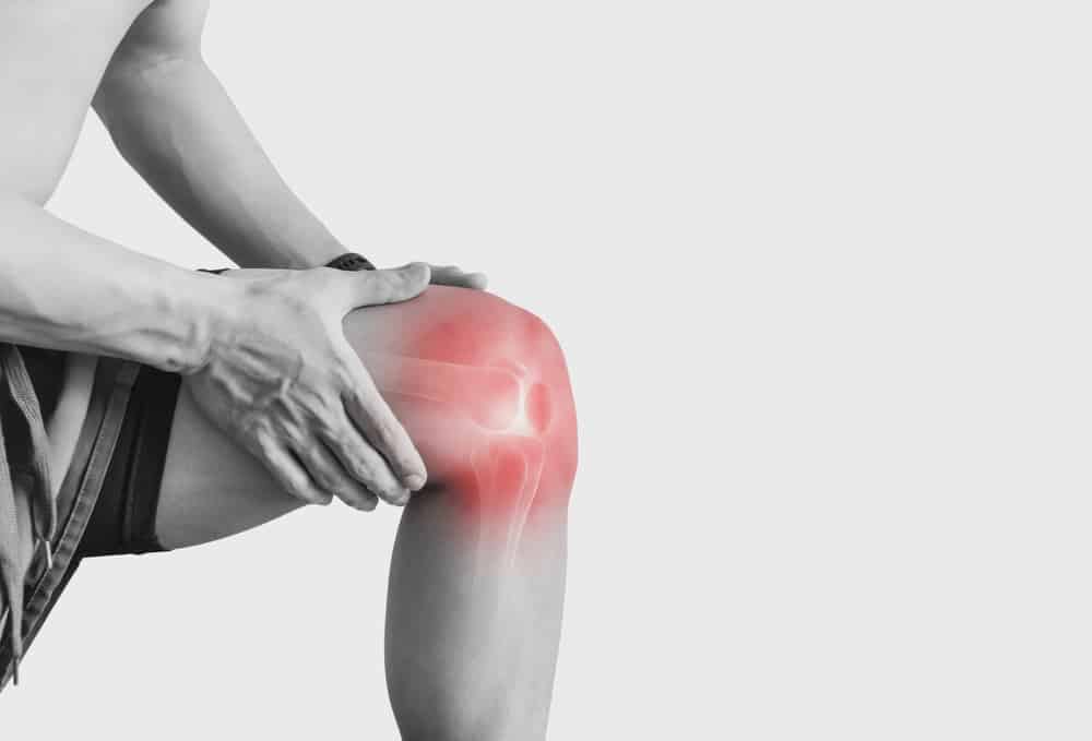
The KT1000 Arthrometer emerges as an essential tool in the field of knee injury diagnosis and management. Renowned for its unparalleled precision in measuring knee laxity, this device has become a staple in orthopedic and sports medicine clinics worldwide. Its ability to accurately gauge the anterior-posterior movement of the tibia relative to the femur provides clinicians with crucial data, particularly in assessing the condition of the knee’s ligaments.
Employed extensively for evaluating the integrity of the anterior cruciate ligament (ACL), the KT1000 stands out not only for its diagnostic accuracy but also for its role in guiding treatment decisions and assessing post-operative recovery. This article aims to illuminate the pivotal role of the KT1000 in contemporary orthopedic practice, underscoring its significance in enhancing patient outcomes in knee-related injuries and disorders.
1. Background and History
The journey of the KT1000 Arthrometer in the medical landscape began as a response to the growing need for more precise and quantitative assessments of knee joint laxity. Developed in the late 20th century, this device marked a significant advancement over the traditional manual examination techniques, which were often subjective and lacked reproducibility.
Initially, the KT1000 arthrometer was conceptualized to provide a standardized method to measure the anterior-posterior displacement of the tibia, particularly crucial in the diagnosis and management of ACL injuries. ACL ruptures are common, especially in athletes, and the accurate assessment of the extent of injury is vital for deciding the course of treatment—be it surgical intervention or conservative management.
Over the years, the KT1000 arthrometer has undergone various enhancements, evolving in its design and functionality to meet the advancing standards of orthopedic care. Its introduction was a turning point, enabling doctors to make more informed decisions based on objective data. The precision of the KT1000 arthrometer provided a reproducible and reliable means to not only diagnose ligament injuries but also to monitor rehabilitation progress post-injury or surgery.
The KT1000’s significance was further cemented by numerous studies and research papers that validated its efficacy and reliability. These studies highlighted its role in improving the diagnostic accuracy for knee ligament injuries, thereby influencing treatment protocols and patient outcomes positively.
Today, the KT1000 arthrometer is not just a diagnostic tool but a symbol of the technological strides made in orthopedics. Its history reflects the continuous pursuit of precision and reliability in medical diagnostics, embodying the progress from subjective assessment to evidence-based, quantitative analysis. This evolution has not only benefitted patients with more accurate diagnoses and tailored treatments but has also set a benchmark for future innovations in the field of orthopedic diagnostic tools.
2. Technical Specifications
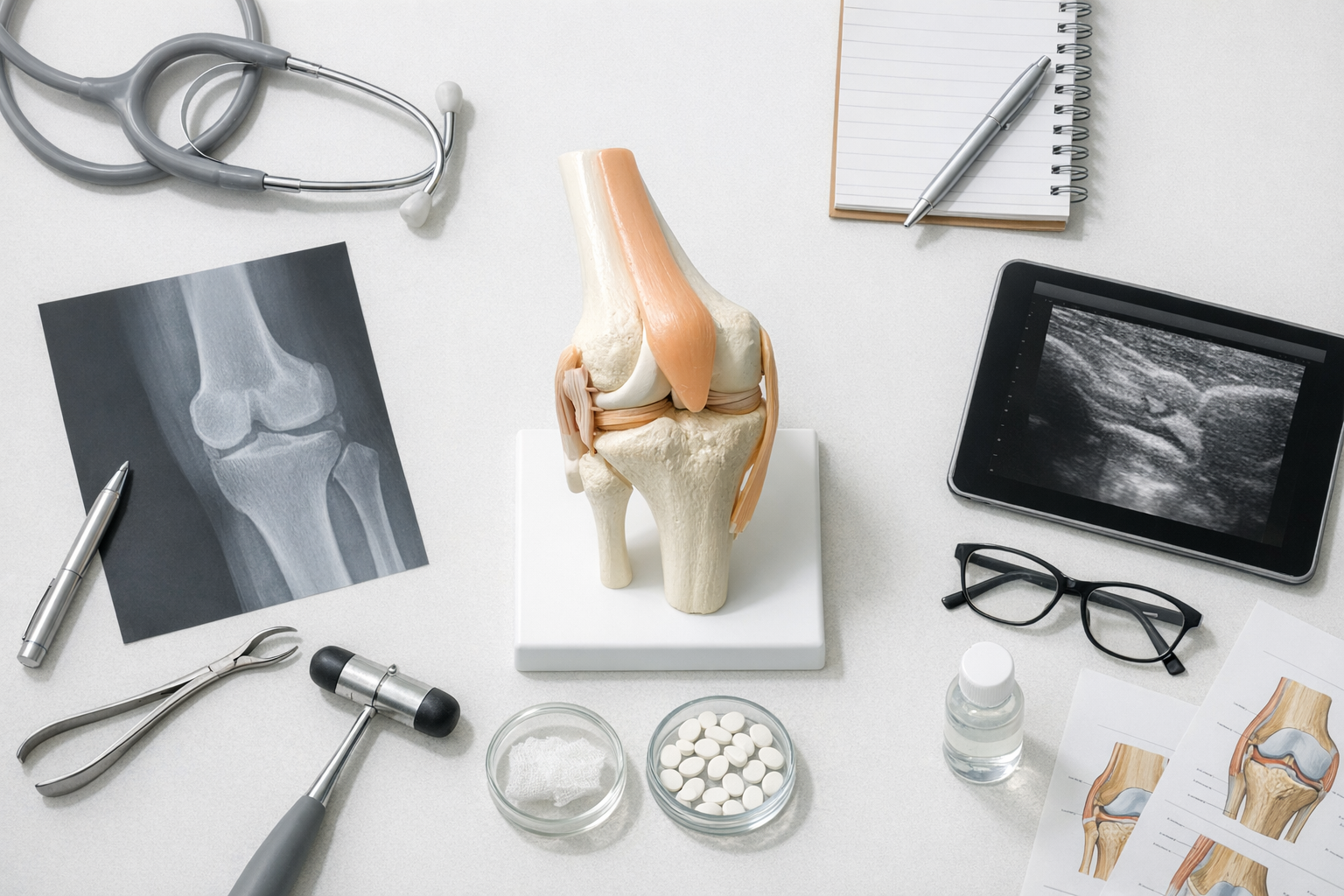
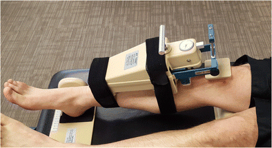
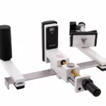
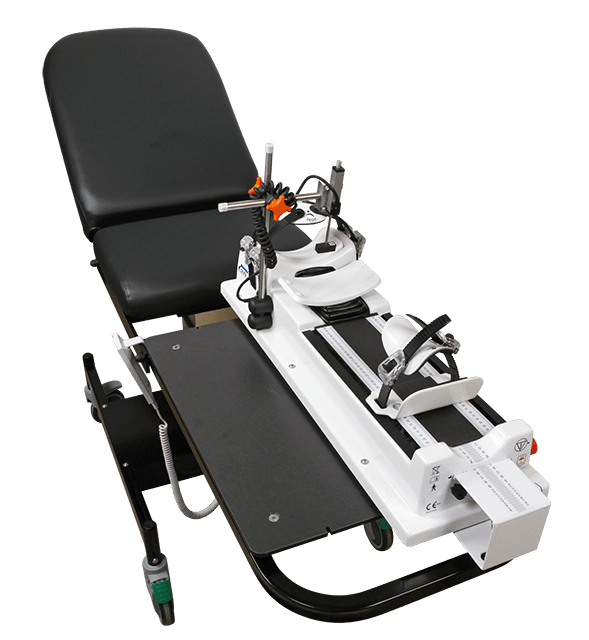
The KT1000 Arthrometer is a hallmark of engineering precision, designed to offer accurate and consistent measurements of knee joint laxity. Central to its functionality is the device’s ability to quantitatively assess the anterior-posterior displacement of the tibia relative to the femur, a key indicator in evaluating ligament integrity, especially the anterior cruciate ligament (ACL).
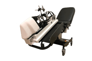
Construction and Design: The KT1000 comprises a main body with a sensor pad, which is placed on the patient’s tibia. The device is equipped with a finely calibrated measuring gauge that records the tibial movement in millimeters. This precision allows for a detailed assessment of the knee’s stability and the extent of ligamentous injury.
Measurement Capability: One of the standout features of the KT1000 arthrometer is its capability to measure displacements ranging from 0 to 30 millimeters, with an accuracy of up to 1 millimeter. This range is crucial for diagnosing varying degrees of ligament laxity, from mild to severe.
Adjustability and Comfort: Designed with patient comfort in mind, the KT1000 arthrometer is adjustable to accommodate different leg sizes and shapes. This flexibility ensures accurate positioning and consistent results, irrespective of the patient’s physique.
Force Application System: The device applies a standardized force to the knee, typically ranging between 15 to 30 pounds, depending on the test protocol. This standardized force application is critical for reproducible results, as it eliminates the variability inherent in manual testing methods.
Digital Output: Advanced models of the KT1000 arthrometer feature digital displays for immediate reading of measurements, enhancing the ease of use for clinicians. This digital integration ensures not only accuracy in readings but also facilitates efficient recording and tracking of patient data over time.
In summary, the technical prowess of the KT1000 lies in its precision, adjustability, and the ability to provide objective, quantifiable data. These specifications make it an indispensable tool in the diagnosis and management of knee injuries, particularly in cases where accurate assessment of ligament integrity is crucial for determining the appropriate treatment plan.
3. Methodology of Testing
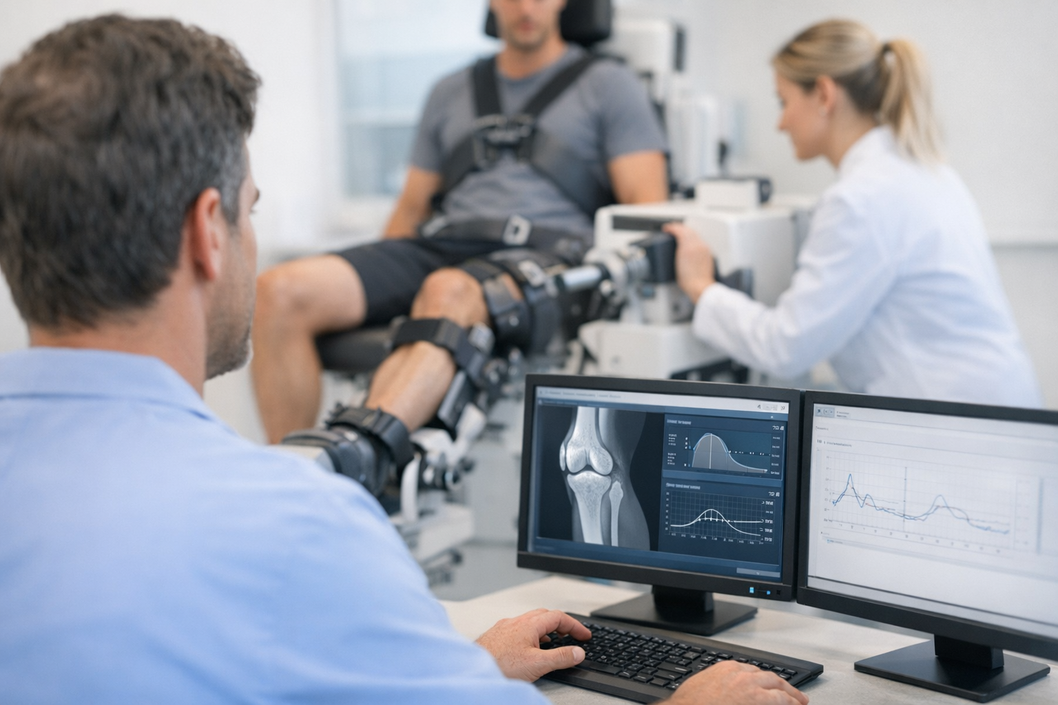
The KT1000 Arthrometer is a sophisticated tool used for assessing knee joint laxity, primarily to evaluate the condition of the anterior cruciate ligament (ACL). Its testing methodology is precise, standardized, and forms a critical part of the diagnostic process in orthopedics and sports medicine. Here’s a detailed breakdown of the testing procedure:
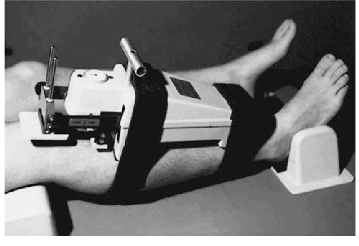
Patient Positioning: The accuracy of KT1000 testing begins with proper patient positioning. The patient is typically seated or lies down with the knee in a slightly bent position (approximately 25 to 30 degrees of flexion). This position relaxes the hamstring muscles and ensures an accurate measurement of tibial movement relative to the femur.
Device Placement: The clinician places the KT1000’s sensor pad on the proximal tibia, ensuring that it’s properly aligned. It’s crucial that the device is positioned consistently for each measurement to ensure reliability and reproducibility of the results.
Baseline Measurement: Before applying any force, a baseline measurement of the tibia’s position is taken. This initial reading is critical as it serves as a reference point against which the subsequent displacement is measured.
Force Application: The KT1000 applies a standardized anterior force to the proximal tibia, simulating the stress placed on the knee ligaments during physical activities. The force applied is typically in the range of 15 to 30 pounds, depending on the test protocol. The clinician may perform the test at different force levels to assess the knee’s response to varying degrees of stress.
Measurement Recording: As the force is applied, the KT1000 arthrometer measures the anterior displacement of the tibia in millimeters. This displacement is a direct indicator of the knee’s anterior-posterior stability and is used to assess the condition of the ACL.
Comparative Assessment: For a comprehensive evaluation, measurements are often taken on both knees. Comparing the results from the injured knee with the uninjured knee provides a clearer picture of the extent of injury and the knee’s overall stability.
Repeatability and Consistency: The test is repeated several times to ensure consistency in the measurements. The repeatability of the results is a testament to the KT1000’s precision and reliability as a diagnostic tool.
Interpreting Results: The clinician interprets the results in the context of the patient’s overall clinical picture, including symptoms, physical examination findings, and other diagnostic tests. A greater degree of tibial displacement indicates higher laxity, which may suggest a compromised ACL.
In summary, the methodology of testing with the KT1000 arthrometer is a well-defined process that requires meticulous attention to detail. From patient positioning to the interpretation of results, each step is crucial in ensuring accurate and reliable assessment of knee laxity. This thorough approach underscores the KT1000’s role as an indispensable tool in the diagnostic arsenal for knee injuries.
4. Clinical Applications

The KT1000 Arthrometer has established itself as an essential diagnostic tool in various clinical settings, particularly in the realms of orthopedics and sports medicine. Its primary application is in the assessment and management of knee injuries, with a special focus on the anterior cruciate ligament (ACL). Here are some key clinical applications of the KT1000:
Diagnosis of ACL Injuries: The KT1000 arthrometer is invaluable in diagnosing ACL injuries, which are common in athletes and can occur during high-impact or twisting movements. By measuring the anterior-posterior displacement of the tibia, the KT1000 helps determine the extent of injury to the ACL, guiding clinicians in deciding between conservative management and surgical intervention.
Post-Operative Assessment: Post-surgical evaluation of ACL reconstruction is another critical area where the KT1000 excels. It assesses the stability of the knee after surgery, helping clinicians evaluate the success of the procedure and guide rehabilitation protocols.
Screening Athletes: In sports medicine, the KT1000 arthrometer is used for pre-season screening of athletes, especially in sports with a high incidence of knee injuries. This screening helps identify pre-existing conditions that may predispose athletes to injuries, enabling preventative strategies and tailored training programs.
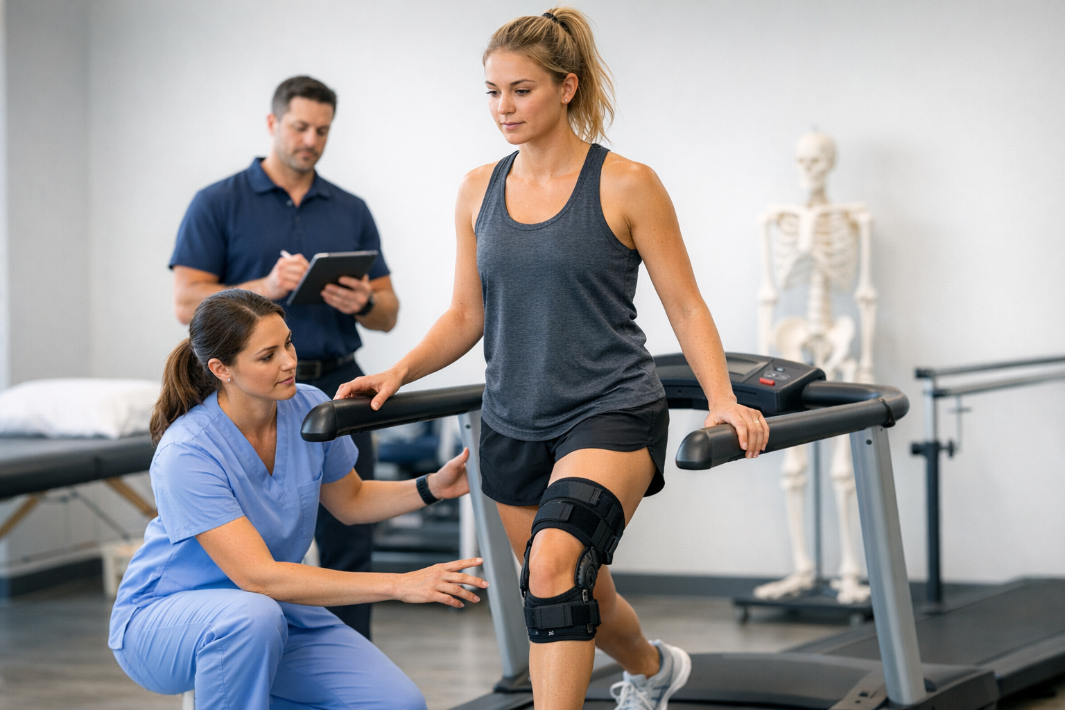
Rehabilitation Monitoring: The device plays a significant role in monitoring the progress of patients undergoing rehabilitation for knee injuries. By providing quantitative data on knee laxity, the KT1000 allows clinicians to track improvements and adjust rehabilitation programs accordingly.
Research Applications: Beyond clinical practice, the KT1000 is widely used in research to study knee biomechanics and the efficacy of various treatment modalities for knee injuries. Its ability to provide objective data makes it a valuable tool in clinical research and in advancing the understanding of knee pathologies.
In essence, the KT1000 Arthrometer is more than just a diagnostic device; it is a versatile tool that aids in the entire continuum of care for knee injuries, from initial assessment to post-operative rehabilitation and beyond. Its clinical applications underscore its significance in enhancing patient outcomes and advancing the field of knee injury management.
5. Comparative Analysis
5.1 GNRB and Dyneelax
Advanced Precision and Consistency: These newer devices, as evidenced in studies (DOI: 10.1016/j.knee.2023.03.017), offer enhanced precision and consistency, crucial for diagnosing partial ACL tears. Studies like Colette et al. & Klasan et al. indicate their superiority in precision compared to the KT units.
Comprehensive Assessments: GNRB® and DYNEELAX® provide more thorough analyses, including rotational laxity (DOI: 10.1016/j.medntd.2023.100254), contributing to a deeper understanding of knee mechanics.
Automated Technology: The GNRB® and DYNEELAX® integrate automation and advanced technology, improving reliability and applicability in various clinical scenarios.
Extension of KT1000 Legacy: While following the foundational principles of the KT1000, these devices represent advancements in arthrometry, combining traditional assessment methods with innovative technology.
Rehabilitation Insights: Studies like Semay et al. (2016) and Nouveau et al. (2017) highlight the role of automated arthrometers in effective rehabilitation monitoring, showcasing their importance in post-surgical recovery processes.
5.2 Telos
The Telos, another arthrometer, although less emphasized in recent studies, has historically played a significant role in knee injury diagnostics. It applies stress Radiography to analyse the knee ligaments.
Invasiveness and Exposure: Stress radiography, involving X-rays, is more invasive and exposes patients to radiation, unlike the non-invasive KT1000.
Functional vs. Imaging Data: KT1000 offers specific functional data on knee laxity, while stress radiography provides visual insights into joint stability. These studies show the superiority of the GNRB compared to the Telos (Lefevre et al. & Bouguennec et al.) in detecting partial ACL thickness tears and reproducibility.
Ease of Use: The KT1000 is more user-friendly and accessible in clinical settings compared to the equipment-intensive and invasive nature of stress radiography.
Complementary Approaches: Despite their differences, both methods are valuable; the KT1000’s functional assessment complements the visual data from stress radiography for a holistic understanding of knee injuries.
Its comparison with GNRB, as explored in Lefevre et al. (2013), reflects the continuous evolution in arthrometry technology.
5.3 MRI
When comparing the KT1000 Arthrometer to MRI in knee injury diagnostics:
- Functional vs. Structural Assessment: The KT1000 focuses on functional assessment of knee laxity, whereas MRI provides detailed structural imaging, crucial for identifying tissue damage.
- Non-Invasiveness: Both KT1000 and MRI are non-invasive, but MRI involves a longer procedure and may require contrast agents.
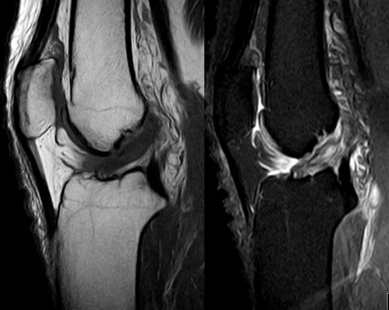
- Diagnostic Scope: MRI offers a comprehensive view of the knee, including soft tissue and bone, while the KT1000 specifically measures ligament laxity. Advanced arthrometers like the GNRB exhibit greater sensitivity in detecting partial ACL tears than MRI (Link to Study ->DOI: 10.1016/j.knee.2023.03.017).
- Clinical Application: While MRI is more comprehensive, the KT1000’s ease of use and specific focus on knee laxity make it a practical choice in many clinical scenarios.
Recent advancements also stress the importance of correlating arthrometer findings with MRI results, as seen in Cojean’s 2023 study (Link To Study -> DOI: 10.1016/j.medntd.2023.100254). MRI provides detailed static analysis, while arthrometers like the KT1000, GNRB® and DYNEELAX® offer dynamic, functional testing, contributing to a more comprehensive diagnostic approach.
6. Challenges and Limitations
The KT1000 Arthrometer, despite its widespread use, faces certain limitations. Operator dependency can lead to variability in results, potentially affecting diagnostic accuracy. Additionally, the manual nature of the device may limit its sensitivity in detecting subtle changes in knee laxity.
Ongoing research and improvements could focus on enhancing its automation to reduce human error. Incorporating digital technology for more precise measurements and data analysis is another area of potential advancement. These enhancements would not only improve diagnostic accuracy but also extend the KT1000’s applicability in diverse clinical settings, ensuring its continued relevance in orthopedic diagnostics.
7. The Future of Arthrometry
The future of arthrometry is poised for significant advancements, integrating technologies like artificial intelligence (AI) and improved sensor accuracy. This evolution will enable more precise, automated measurements and sophisticated data analysis.
Innovations in products like GNRB® and DYNEELAX® are indicative of this forward momentum, emphasizing the industry’s dedication to evolving diagnostic tools. The continual advancement in arthrometry underscores its vital role in adapting to the dynamic demands of modern medical diagnostics.
Conclusion
The KT1000 Arthrometer remains a cornerstone in orthopedic diagnostics for knee injuries. Despite emerging technologies and newer models like GNRB® and DYNEELAX®, the KT1000’s foundational principles continue to influence current and future innovations in arthrometry. Its importance in clinical practice, particularly in assessing knee laxity and guiding treatment decisions, is undisputed. As the field of arthrometry evolves, the KT1000’s legacy endures, underscoring its enduring significance in medical diagnostics.
Medical References
Collette, M., Courville, J., Forton, M., & Gagnière, B. (2012). Objective evaluation of anterior knee laxity; comparison of the KT-1000 and GNRB arthrometers. Knee Surg Sports Traumatol Arthrosc. DOI: 10.1007/s00167-011-1869-2.
Lefevre, N., Bohu, Y., Naouri, J. F., Klouche, S., & Herman, S. (2013). Validity of GNRB Arthrometer Compared to Telos in the Assessment of Partial Anterior Cruciate Ligament Tears. Knee Surg Sports Traumatol Arthrosc. DOI: 10.1007/s00167-013-2384-4.
Bouguennec, N., Odri, G.A., Graveleau, N., & Colombet, P. (2015). Comparative reproducibility of TELOS™ and GNRB® for instrumental measurement of anterior tibial translation in normal knees. Orthopaedics & Traumatology: Surgery & Research. DOI: 10.1016/j.otsr.2015.01.007.
Senioris, A., Rousseau, T., L’Hermette, M., Gouzy, S., Duparc, F., & Dujardin, F. (2017). Validity of rotational laxity coupled with anterior translation of the knee. The Knee. DOI: 10.1016/j.knee.2017.01.009.
Ryu, S. M., Na, H. D., & Shon, O. J. (2018). Diagnostic Tools for Acute Anterior Cruciate Ligament Injury: GNRB Lachman Test and Telos. Knee Surg Relat Res. DOI: 10.5792/ksrr.17.014.
Klasan, A., Putnis, S.E., Kandhari, V., Oshima, T., & Parker, D.A. (2019). Anterior knee translation measurements after ACL reconstruction are influenced by the type of laximeter used. Knee Surgery Sports Traumatology Arthroscopy. DOI: 10.1007/s00167-020-05950-5.
Forelli, F., Le Coroller, N., Gaspar, M., Memain, G., Kakavas, G., Miraglia, N., Marine, P., Maille, P., Hewett, T. E., & Rambaud, A. J. M. (2023). Ecological and Specific Evidence-Based Safe Return To Play After Anterior Cruciate Ligament Reconstruction In Soccer Players: A New International Paradigm. International Journal of Sports Physical Therapy. DOI: 10.26603/001c.73031.
Cojean, T., Batailler, C., Robert, H., & Cheze, L. (2023). GNRB® laximeter with magnetic resonance imaging in clinical practice for complete and partial anterior cruciate ligament tears detection: A prospective diagnostic study with arthroscopic validation on 214 patients. The Knee. DOI: 10.1016/j.knee.2023.03.017.
Cojean, T., Batailler, C., Robert, H., & Cheze, L. (2023). Sensitivity, repeatability, and reproducibility study with a leg prototype of a recently developed knee arthrometer Dyneelax. Medicine in Novel Technology and Devices, 19, 100254. DOI: 10.1016/j.medntd.2023.100254.
Nouveau, S., Robert, H., & Viel, T. (2017). ACL Grafts Compliance During Time: Influence of Early Solicitations on the Final Stiffness of the Graft after Surgery. Journal of Orthopedic Research and Physiotherapy, 3(1), 035. DOI: 10.24966/ORP-2052/100035.