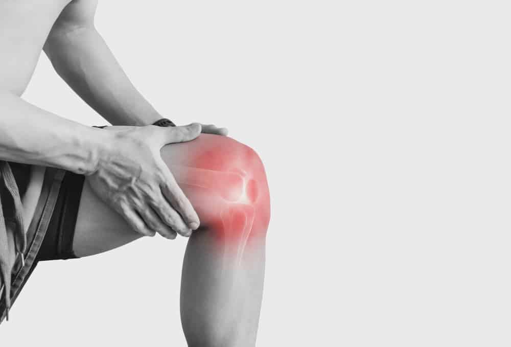
In the first hours and days after an ACL tear, examination findings can shift quickly. That is why acute ACL injury timing matters: swelling, pain, hemarthrosis, and reflex muscle contraction may all change what the clinician feels and what the patient tolerates. For orthopaedic surgeons, sports physicians, physiotherapists, and ER teams, understanding early ACL assessment is less about finding one perfect test and more about interpreting findings in context. The practical question is not only whether the ACL is injured, but also how acute ACL injury timing affects laxity, rotational findings, confidence in diagnosis, and the need for re-examination, imaging, or urgent referral.
1. Why acute ACL injury timing changes the exam
The earliest assessment after a suspected ACL tear is often performed in a knee that is painful, swollen, and guarded. In that setting, acute ACL injury timing can alter both the sensitivity of bedside tests and the examiner’s confidence in the result. This is especially relevant for timing of ACL diagnosis in urgent care, emergency, and sideline-to-clinic pathways.
Several mechanisms explain the problem:
- Hemarthrosis increases intra-articular pressure and limits motion.
- Pain reduces relaxation and can make the knee difficult to position.
- Muscle guarding after ACL tear, especially hamstring co-contraction, may resist anterior tibial translation.
- Associated injuries such as meniscal tears, MCL injury, bone bruising, or posterolateral pathology can further distort the exam.
In practical terms, acute ACL injury timing means that a negative or uncertain early test does not reliably exclude clinically important instability. This is one reason repeat examination, structured follow-up, and selective imaging are often necessary. Early timing also affects triage and referral decisions in a triage algorithm.
Some newer imaging work also reinforces that ACL injury is not simply a binary torn-versus-intact issue. For example, Li et al. (2026) evaluated quantitative MRI during hyperextension and explored abnormalities in the screw-home mechanism after ACL injury, reminding clinicians that altered knee kinematics may extend beyond what a routine static snapshot captures. That does not change the basic acute bedside approach, but it supports the idea that acute ACL injury timing can affect what is visible functionally versus structurally.
2. acute ACL injury timing and the first 72 hours
During the first 24 to 72 hours, the biggest pitfalls are swelling, pain, and protective muscle contraction. If the patient reports a pop, rapid swelling, giving way, and inability to continue activity, suspicion may be high even when manual testing is equivocal. This is where ACL examination timing becomes clinically important.
2.1 The effect of hemarthrosis
hemarthrosis and ACL exam are tightly linked. A tense effusion may limit extension, increase pain with motion, and make the patient apprehensive before the examiner even reaches the endpoint of a test. The result can be a falsely reassuring examination or one that is simply uninterpretable.
In these acute presentations, a careful history plus inspection, aspiration when clinically indicated and appropriate, neurovascular review, fracture exclusion, and short-interval reassessment may be more useful than over-interpreting one painful maneuver. This is also why broad concepts of knee laxity matter: what appears mechanically stable in the first visit may be partly a function of swelling and guarding rather than true ligament integrity.
2.2 The effect of guarding
muscle guarding after ACL tear can significantly reduce perceived anterior tibial movement. Hamstring activation is especially important because it opposes anterior translation of the tibia. If the patient is bracing, grimacing, or unable to relax, the examiner may feel a firmer stop than expected.
This is one reason acute knee laxity testing is often less straightforward than in the subacute clinic setting. The relationship between hamstring stiffness and translation is discussed further in hamstring stiffness, and it is directly relevant when interpreting why two examinations performed days apart may appear different despite the same underlying ACL injury.
A short decision aid can help:
- If pain and effusion are high, avoid forcing endpoint-based interpretation.
- If suspicion remains high despite equivocal testing, arrange re-examination after swelling and guarding settle.
- Use MRI to assess associated injuries and structural detail when clinically indicated.
- Escalate sooner if there is locked knee, gross instability, fracture concern, neurovascular symptoms, or multi-ligament suspicion.
3. Which tests change most with acute ACL injury timing
Not all tests are equally affected by acute ACL injury timing. The challenge is to know which findings remain useful early, which become less reliable, and which may be best deferred or repeated.
3.1 Lachman in the acute setting
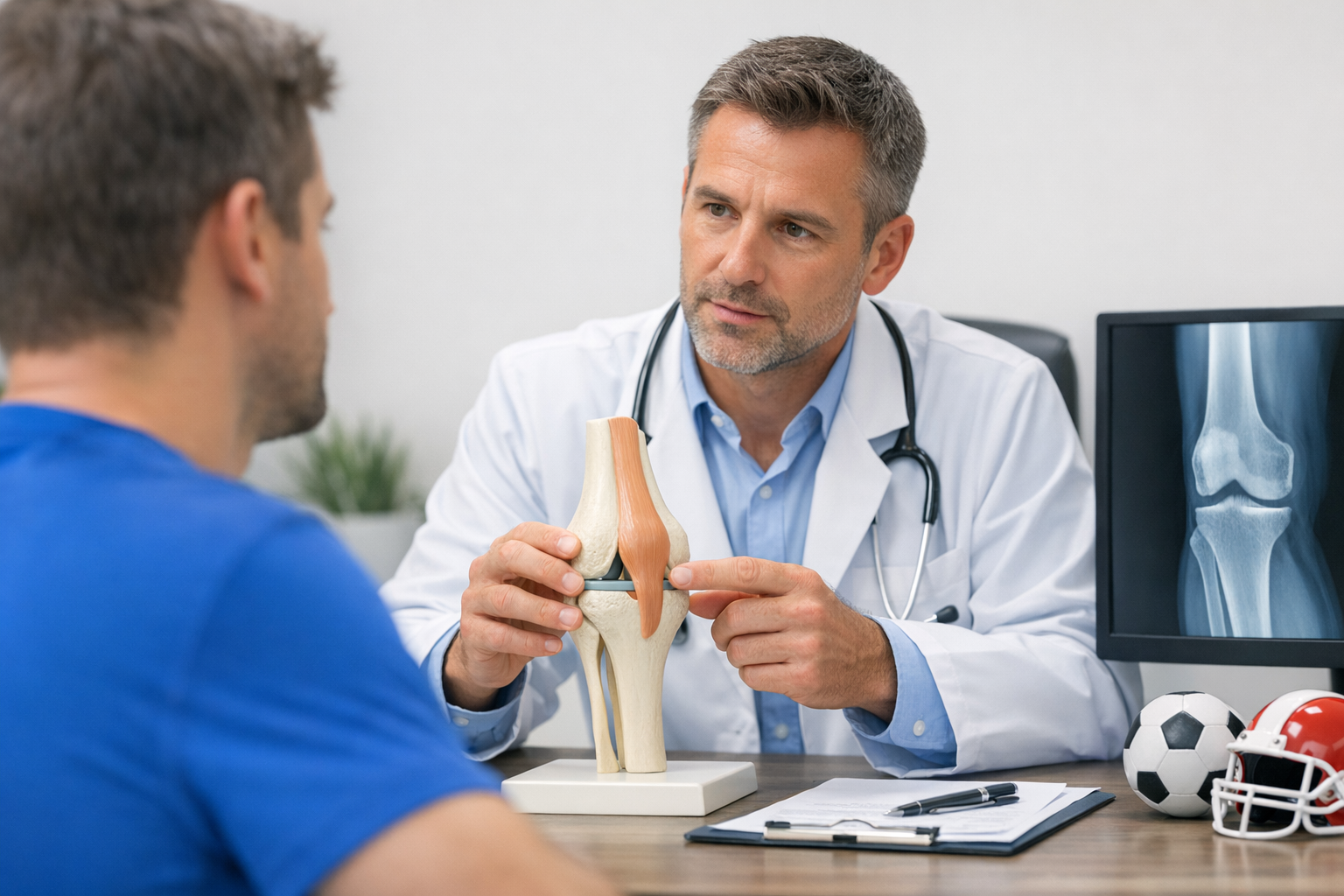
The Lachman test acute ACL assessment is often the most useful manual test early on, but it still depends on relaxation and examiner skill. A soft endpoint or increased translation compared with the other side may support ACL disruption. However, severe pain, large effusion, or poor positioning may reduce confidence.
For a deeper breakdown of technique and limitations, see the Lachman test guide. In acute presentations, the main pitfall is not only false negatives, but also overconfidence in a limited exam. This is why acute ACL injury timing should always be documented alongside the test result.
3.2 Anterior drawer and pivoting tests
The anterior drawer may be less helpful acutely if flexion is restricted or the hamstrings are active. Meniscal blockage and patient apprehension can also interfere. Broader comparisons between test options are reviewed in diagnostic tests.
The most timing-sensitive maneuver is often the rotational exam. pivot shift in acute ACL injury may be difficult or impossible to elicit because it is painful, guarding is high, and the patient may not tolerate the combined valgus and internal rotation load. A negative pivot shift very early does not rule out significant rotational instability.
That point matters because rotational control can be central to functional symptoms. When the exam is repeated after swelling subsides, the same knee may move from an equivocal to a clearly positive finding. In the rare but important setting of combined injury patterns, rotational findings may also be distorted by associated ligament damage, as illustrated by Shahare et al. (2026), who described an unusual bilateral posterolateral corner injury presentation with asymmetric cruciate tears.
4. When early diagnosis is uncertain
Sometimes the first visit produces a high-suspicion story with low-confidence physical findings. That is where acute ACL injury timing becomes a management issue rather than just a diagnostic nuance. The right next step depends on what uncertainty remains.
Common reasons for early uncertainty include:
- Pain-limited examination
- Large effusion or loss of extension
- Borderline translation on Lachman
- No clear pivot shift because of guarding
- Concern for partial tear or associated ligament injury
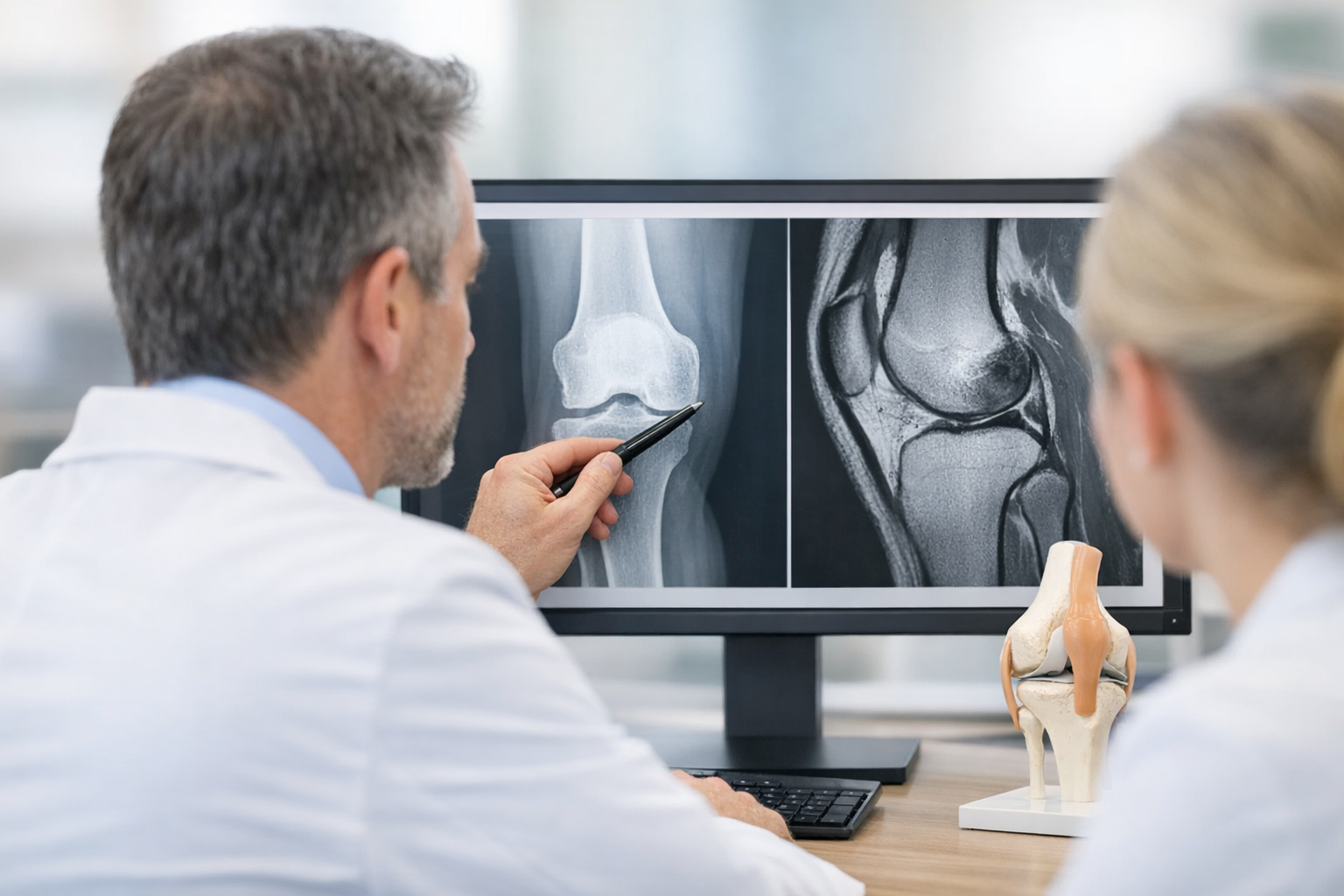
In these cases, MRI remains important because it evaluates meniscus, cartilage, bone bruising, and collateral or posterolateral structures. At the same time, MRI does not directly quantify dynamic side-to-side instability. A balanced framework is outlined in MRI vs arthrometer, where imaging and functional assessment are positioned as complementary rather than competing tools.
If MRI is equivocal or a partial tear is suspected, objective laxity testing may help quantify functional instability that static imaging may not fully capture, although MRI typically remains necessary to assess associated injuries and to support pre-operative planning when reconstruction is considered. That issue is particularly relevant in borderline MRI workflows.
For broader context on case finding across settings, see this overview of ACL detection. In day-to-day practice, acute ACL injury timing should push clinicians away from false certainty and toward staged interpretation.
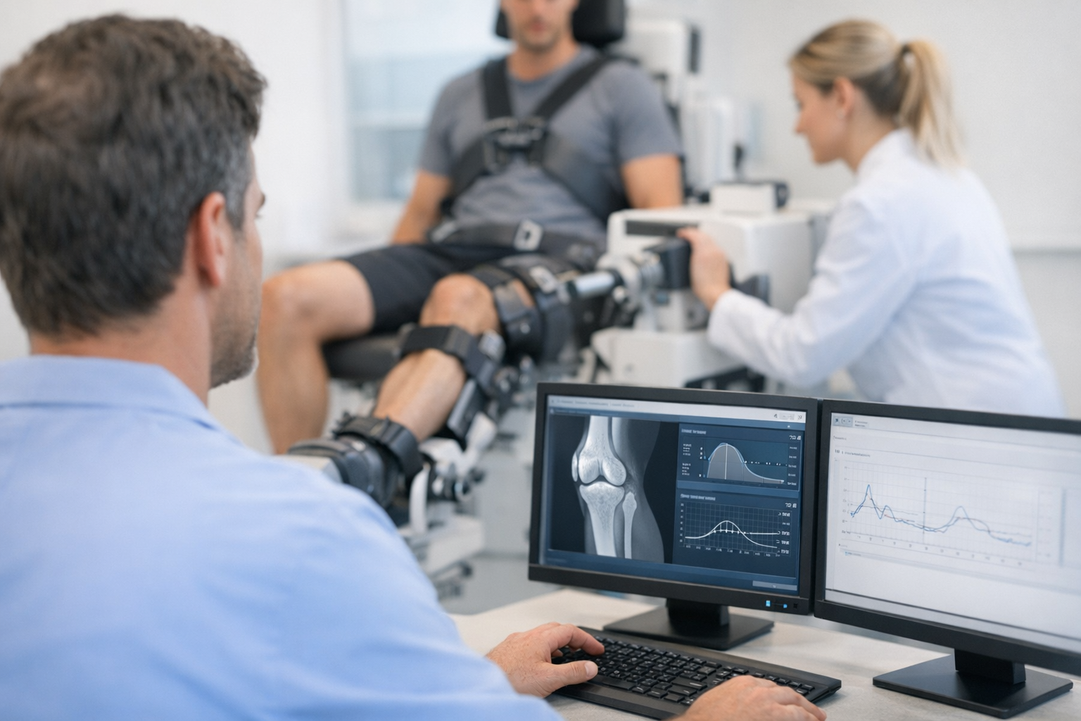
5. Where objective laxity testing may fit
When repeated manual examination remains limited or when documenting side-to-side anterior instability matters, objective laxity testing may complement the clinical exam by quantifying translation over controlled loading conditions, and in some pathways devices such as the GNRB arthrometer or Dyneelax may add useful functional information alongside MRI rather than replacing it.
This is particularly relevant because acute ACL injury timing can make side-to-side comparison difficult by hand alone. Objective testing may be most informative after the patient can tolerate positioning and when guarding is reduced enough for interpretable measurement. It may also help in follow-up when the clinician is tracking whether apparent instability changes are due to true laxity versus early pain inhibition.
That said, these tools still require clinical judgment. Poor tolerance, severe swelling, or multi-ligament injury may limit interpretation. They are best used as part of a clinician-led pathway, not as standalone diagnostic proof.
6. Why timing matters beyond diagnosis
acute ACL injury timing does not only affect whether the tear is detected. It also influences downstream decisions about activity restriction, rehabilitation start points, MRI prioritization, and referral urgency. The consequences of delayed recognition may include recurrent giving way, secondary meniscal injury, and progressive cartilage stress.
Although the studies provided here are not direct diagnostic accuracy trials for acute bedside testing, they support the broader importance of timely recognition and knee environment after ACL rupture. Jin et al. (2025) analyzed risk factors for cartilage damage in patients with ACL rupture. Lee et al. (2026) examined how body mass index influences tibiofemoral cartilage composition and biochemistry after ACL injury. In an experimental model, Retzky et al. (2026) suggested that early reconstruction may mitigate posttraumatic osteoarthritis development. These studies should not be overextended into a rigid acute treatment rule, but they do reinforce a practical message: timing of ACL diagnosis matters because post-injury mechanics and joint health evolve.
This is also relevant in younger patients, where reassessment and longitudinal monitoring may be especially important. For that perspective, see pediatric ACL follow-up considerations.
A useful clinical summary is:
- Very early exam: best for suspicion, triage, and red-flag detection.
- Early follow-up exam: often better for interpreting laxity and endpoint quality.
- MRI: helps define structure and associated injury burden.
- Functional laxity assessment: may clarify instability when manual findings are limited or borderline.
7. Key takeaways and next steps
acute ACL injury timing should be treated as a core part of interpretation, not just a background detail. The same knee may appear different at 6 hours, 3 days, and 2 weeks because pain, swelling, and guarding change what the examiner can feel. This is why early ACL assessment is often provisional rather than final.
The practical takeaway is simple:
- Do not over-reassure based on one painful negative exam.
- Use history, swelling pattern, function, and comparative testing together.
- Recognize that hemarthrosis and ACL exam findings are closely linked.
- Expect pivot shift in acute ACL injury to be less reliable early.
- Re-examine when needed, and use MRI and other complementary tools thoughtfully.
For clinicians, the safest approach is staged assessment with clear documentation of symptoms, exam limits, and reassessment plans. In other words, acute ACL injury timing is not a minor variable. It is often the reason findings change, confidence shifts, and the diagnostic pathway needs to be repeated or refined.
Clinical references (PubMed)
1) 2026 – Li et al. – Quantitative magnetic resonance imaging during knee hyperextension for the evaluation of screw-home mechanism abnormalities in patients with anterior cruciate ligament injury: a clinical study. – Quant Imaging Med Surg – DOI: 10.21037/qims-2025-1-2628 – PMID: 42273143 – PubMed
2) 2025 – Jin et al. – Analysis of risk factors for knee cartilage damage in patients with anterior cruciate ligament rupture. – Medicine (Baltimore) – DOI: 10.1097/MD.0000000000046103 – PMID: 41431029 – PubMed
3) 2026 – Retzky et al. – Early Anterior Cruciate Ligament Reconstruction Mitigates the Development of Posttraumatic Osteoarthritis in a Murine Anterior Cruciate Ligament Rupture Model. – Am J Sports Med – DOI: 10.1177/03635465251390541 – PMID: 41476416 – PubMed
4) 2026 – Shahare et al. – A Unique Presentation of Bilateral Posterolateral Corner Injury With Right Posterior Cruciate Ligament and Left Anterior Cruciate Ligament Tears. – Cureus – DOI: 10.7759/cureus.106046 – PMID: 42058347 – PubMed
5) 2026 – Lee et al. – Body Mass Index Influences Tibiofemoral Cartilage Composition and Biochemistry in Individuals With Anterior Cruciate Ligament Injury. – J Athl Train – DOI: 10.4085/1062-6050-0027.25 – PMID: 41938330 – PubMed