
ACL injury in tennis is often discussed less than in football or skiing, yet the sport creates a distinct mix of risk through abrupt deceleration, open-stance loading, recovery steps, and occasional sliding on hard or clay courts. A tennis ACL tear may not always follow a collision. Instead, symptoms can begin during a wide forehand, emergency direction change, or unstable landing after an overhead. For clinicians, the challenge is not only diagnosing structural injury, but also identifying rotational control deficits, functional instability, and sport-specific demands that affect treatment and return to sport ACL tennis decisions.
1. Why ACL injury in tennis deserves sport-specific attention
ACL injury in tennis is not simply a generic noncontact knee injury occurring on a tennis court. The sport combines repeated split-step landings, lateral acceleration, trunk rotation, low-base recovery positions, and variable surfaces. These features can expose the knee to multiplanar loading, especially when the foot is planted and the athlete is trying to change direction under time pressure.
In practice, cutting and ACL injury are closely linked in tennis because players often decelerate in a semi-flexed position before pushing laterally or recovering diagonally. That movement is then complicated by racket preparation, visual tracking, and asymmetrical limb use. On clay, tennis sliding knee injury patterns may also involve uncontrolled rotation if the slide starts late, the shoe catches, or the center of mass drifts behind the base of support.
For broader sport-specific context, see this tennis guide.
Recent conceptual work such as Souryal et al. (2026) encourages clinicians to think beyond a single catastrophic moment and consider progressive insufficiency, loading history, and tissue tolerance. That framing is relevant to tennis, where repetitive training volume, fatigue, and cumulative mechanical exposure may contribute to a vulnerable knee before the final giving-way episode occurs.
2. Mechanisms: cutting, sliding, and rotational load in ACL injury in tennis
When evaluating ACL injury in tennis, it helps to separate the movement patterns most likely to overload the ligament.
2.1 Cutting and recovery steps
A classic mechanism involves a player moving wide, planting the foot, and attempting an immediate push-off for recovery. If hip control is delayed, trunk lean is poorly managed, or the knee drifts into valgus and internal rotation, cutting and ACL injury can occur even without contact.
Clinically, ask whether the athlete felt a pop during a directional change, whether the knee shifted during the first step back to center, and whether instability appeared immediately or after swelling developed.
2.2 Sliding injuries
Tennis sliding knee injury is especially relevant on clay but can also occur on hard courts during defensive reach patterns. A controlled slide may reduce peak load in some situations, but a mistimed or asymmetric slide can create abrupt tibial rotation or a delayed catch of the foot. That may stress the ACL, meniscus, anterolateral structures, or MCL depending on the exact position of the knee and trunk.
2.3 Hyperextension and combined injury patterns
Not all tennis presentations are straightforward valgus-rotation events. Hyperextension with capsuloligamentous involvement may signal a more complex pattern. Deardurff et al. (2026) showed in a robotic cadaveric model that injuries to multiple ligamentous and capsular structures can produce abnormal knee hyperextension. In a tennis player with hyperextension, bruising, unusual recurvatum, or severe instability, clinicians should consider whether there is more than an isolated ACL injury.
This matters because management decisions for ACL injury in tennis may change significantly when combined injury or capsular damage is present.
3. Clinical exam: when rotational instability matters most
The initial question is not just whether the ACL is torn, but whether the athlete has functional instability that matches the symptoms. Tennis players often describe the knee as unreliable during braking, turning, or recovering from a stretched position rather than during straight-line activity.
A practical assessment starts with history, effusion, gait, range of motion, and standard ligament testing. The Lachman remains a core exam maneuver in suspected ACL disruption. However, for ACL injury in tennis, clinicians should also pay close attention to rotational findings because tennis movement is rarely purely sagittal-plane.
3.1 Pivot shift and tennis-specific symptoms
Pivot shift in ACL injury is particularly relevant when the athlete reports giving way during a cut, crossover recovery step, or unstable slide. A high-grade pivot shift may suggest substantial rotational instability and can influence both treatment planning and rehabilitation progression. For a deeper discussion, review pivot shift.
Key symptom clues that should raise concern include:
- Giving way during lateral push-off
- Instability when changing direction after a wide ball
- Apprehension during open-stance forehands
- Loss of confidence in sliding or recovery movement
These complaints often align better with rotational instability than with isolated anterior translation alone. That is why ACL instability assessment in tennis should not stop at a binary torn-versus-intact framework.
In cases where MRI findings seem limited but instability symptoms are prominent, a borderline MRI workflow can be clinically useful. The goal is not to dismiss imaging, but to reconcile symptoms, exam findings, and functional testing.
4. ACL instability assessment: how to combine exam, MRI, and objective laxity testing
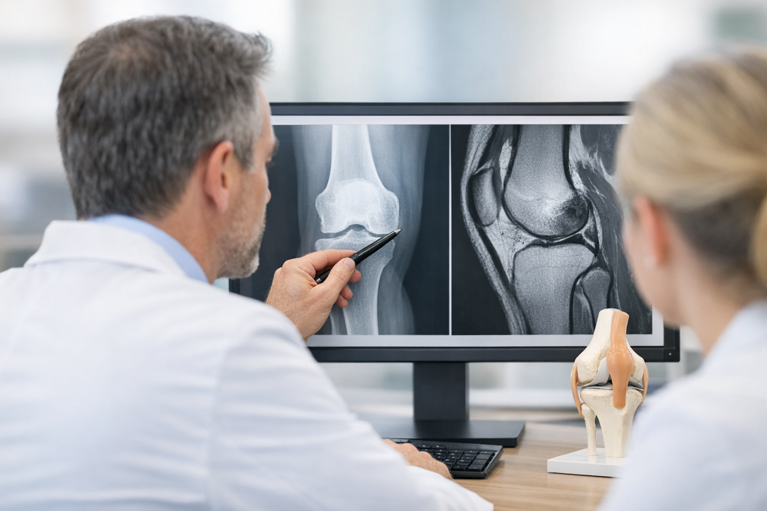
ACL instability assessment in tennis works best when clinicians integrate three domains: structural imaging, hands-on examination, and objective measurement of laxity or dynamic response. MRI remains important for associated meniscal, chondral, osseous, and preoperative information. Still, imaging does not always explain why one player feels unstable during cutting while another with a similar MRI does not.
This is where understanding knee laxity becomes helpful. Subjective instability and measured laxity are related but not identical. Some players report strong instability with modest static laxity because rotational control, neuromuscular timing, pain inhibition, or associated soft tissue injury is the main problem.
A useful clinician-led decision aid for suspected ACL injury in tennis is:
- Confirm injury mechanism, swelling timeline, and instability episodes.
- Perform Lachman, pivot shift when appropriate, collateral and posterolateral examination.
- Use MRI to define ligament integrity and associated intra-articular injury.
- If symptoms and imaging do not align, consider objective knee laxity testing to quantify side-to-side difference and dynamic instability.
- Reassess sport demands, including cutting, deceleration, and sliding exposure.
For an overview of structured evaluation, see this objective knee examination framework. If the question is whether imaging and quantified instability can complement each other, this discussion of MRI vs arthrometer is also relevant.
5. Where instrumented laxity testing fits in tennis ACL evaluation
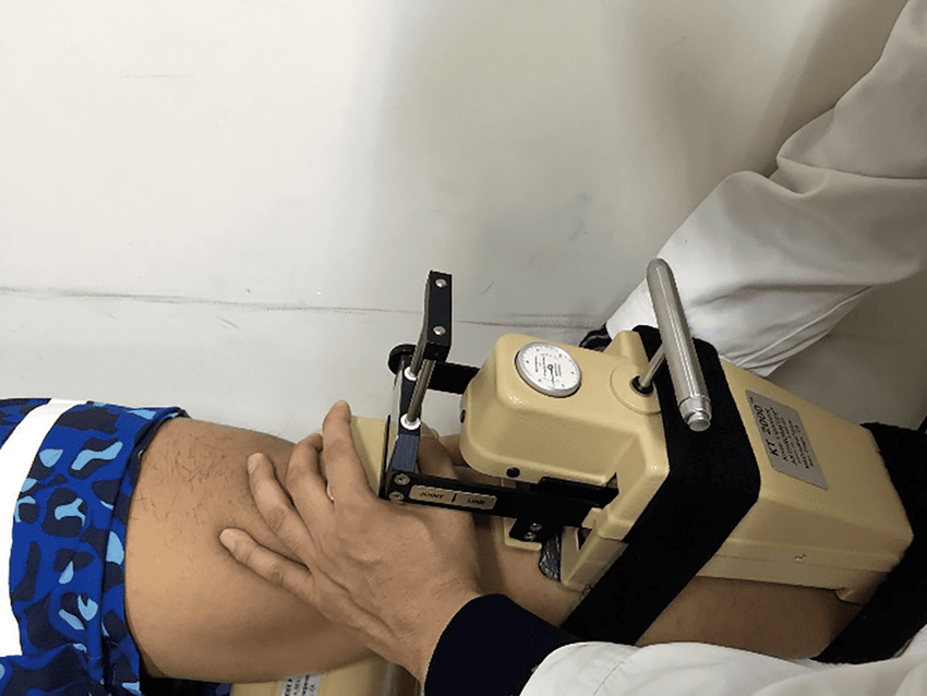
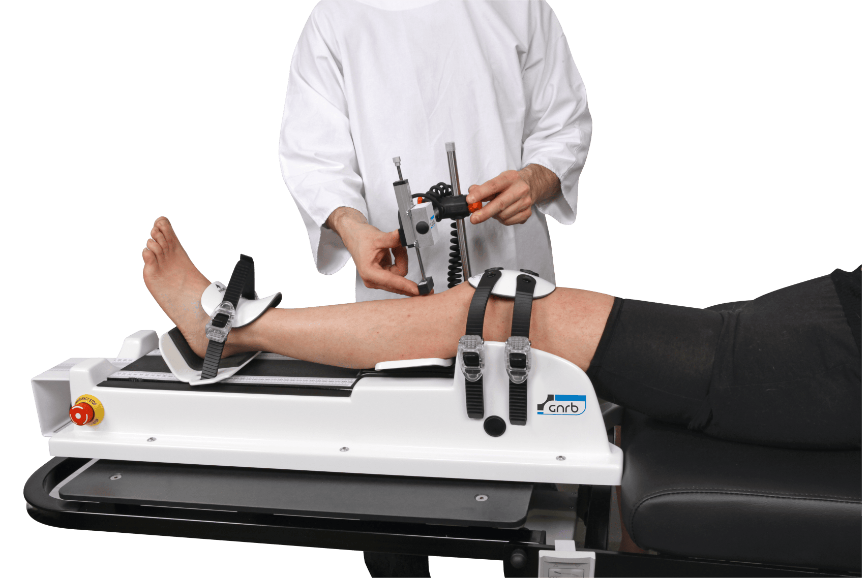
In ACL injury in tennis, clinicians often need more than a descriptive exam note saying the knee feels loose. Objective knee laxity testing can complement the clinical exam and MRI by quantifying side-to-side anterior instability and, in some workflows, adding information about dynamic or multi-axis behavior that may matter in players who cut, brake, and slide repeatedly.
An instrumented knee arthrometer may be especially helpful when:
- Symptoms are disproportionate to MRI findings
- A partial ACL tear is suspected
- There is concern about residual laxity after reconstruction
- Return-to-sport decisions require more objective stability data
This should be framed cautiously. Instrumented assessment does not replace MRI. Rather, it adds functional measurement that imaging may not fully capture, especially in equivocal or borderline presentations. MRI is still typically needed to assess meniscus, cartilage, bone bruising, and other associated pathology, and for surgical planning when reconstruction is considered.
For clinics using objective measurement tools, a GNRB arthrometer can help quantify anterior tibial translation under controlled loading, while a Dyneelax knee arthrometer may fit workflows that include broader dynamic laxity analysis.
Where rotational symptoms are prominent, a dynamic workflow may be more informative than a purely static approach. That distinction matters in ACL injury in tennis because a player may cope during straight-line tasks yet still fail during pivoting or slide-to-recovery actions.
6. Return to sport ACL tennis: what should actually guide clearance?
Return to sport ACL tennis should not be based on time alone. Tennis places high demands on deceleration, unplanned movement, rotational tolerance, repeated low-base loading, and confidence under fatigue. A player who jogs well and passes simple hop tasks may still struggle with live directional demands.
Liang et al. (2026) addressed return-to-play considerations after ACL reconstruction in the APKASS 2024 consensus statement. Although not tennis-specific, the broader message is highly relevant: clearance should be criterion-based, clinically reasoned, and individualized.
For ACL injury in tennis, the decision usually works best when these domains are all considered:
- Symptoms – no instability episodes, manageable pain, controlled swelling
- Clinical exam – acceptable translation and rotational findings
- Strength and performance – sport-relevant force production and movement quality
- Objective stability – when available, quantified laxity data that fits the clinical picture
- Tennis-specific readiness – cutting, emergency recovery, open-stance loading, and sliding where relevant
This is where rehab and stability data should be interpreted together, not in isolation. See rehab milestones for that connection, and review return to sport criteria focused on objective stability.
Clinicians should also remember that tissue behavior and injury prevention may involve more than movement retraining alone. Nyland et al. (2026) discussed fatigue-related ACL injury prevention from a connective tissue perspective, which may be relevant when considering dense tennis calendars, repeated loading, and inadequate recovery.
In high-level athletes, combined injuries also deserve perspective. Although based on soccer and rugby rather than tennis, Jones et al. (2026) reported no difference in career longevity versus matched uninjured controls after ACL reconstruction for combined ACL/MCL injuries in professional players. The exact lesson should not be overgeneralized to tennis, but it supports a more nuanced conversation about prognosis when associated ligament injury is present.
7. Key takeaways and next steps
ACL injury in tennis should be approached as a sport-specific instability problem, not just a generic ligament diagnosis. The combination of cutting, braking, rotation, and sliding means that rotational control and symptom-provoking movement patterns are central to evaluation.
For day-to-day practice, the main points are:
- ACL injury in tennis commonly involves noncontact deceleration and direction change
- Pivot shift in ACL injury may be especially relevant when instability occurs during cutting or slide recovery
- ACL instability assessment should combine history, exam, and MRI
- Objective knee laxity testing can complement MRI when symptoms and imaging do not fully match
- Instrumented knee arthrometer data may help quantify side-to-side instability and support rehabilitation or return-to-play reasoning
- Return to sport ACL tennis decisions should be criterion-based and clinician-led
In short, ACL injury in tennis is best understood through the interaction of mechanism, rotational instability, measured laxity, and sport-specific demand. If a player reports recurrent giving way despite reassuring imaging, or if post-reconstruction progress seems inconsistent with on-court symptoms, reassessing with a structured, objective pathway may clarify the next step.
Clinical references (PubMed)
1) 2026 – Liang et al. – APKASS 2024 consensus statement on anterior cruciate ligament reconstruction, part Ⅲ: Return to play after anterior cruciate ligament reconstruction. – Asia Pac J Sports Med Arthrosc Rehabil Technol – DOI: 10.1016/j.asmart.2026.05.006 – PMID: 42205141 – PubMed
2) 2026 – Deardurff et al. – Injuries to Multiple Ligamentous and Capsular Structures Cause Abnormal Knee Hyperextension: A Robotic Study in Human Knees. – Arthroscopy – DOI: 10.1002/arj.70319 – PMID: 42200688 – PubMed
3) 2026 – Souryal et al. – The Insufficient Anterior Cruciate Ligament: Reframing the Understanding of Noncontact ACL Injuries. – Sports Health – DOI: 10.1177/19417381261451827 – PMID: 42200574 – PubMed
4) 2026 – Nyland et al. – Non-Contact, Mechanical Fatigue-Related ACL Injury Prevention Through Extracellular Matrix Crosslink Preservation: A Narrative Review. – J Funct Morphol Kinesiol – DOI: 10.3390/jfmk11020180 – PMID: 42200886 – PubMed
5) 2026 – Jones et al. – ACL Reconstruction for Combined ACL/MCL Injuries in Professional Soccer and Rugby Players: No Difference in Career Longevity Compared to Uninjured Matched Controls. – Am J Sports Med – DOI: 10.1177/03635465261448234 – PMID: 42200652 – PubMed