ACL injury in female athletes requires a slightly different clinical lens than generic ACL pathways. The core principles of diagnosis remain the same, but female athlete ACL assessment often needs closer attention to generalized laxity, movement strategy, rotational instability, psychological readiness, and sport-specific exposure. For sports physicians, physiotherapists, surgeons, and researchers, the key question is not only whether the ACL is injured, but how instability behaves over time and how that should influence rehabilitation and return-to-sport decisions. This guide summarizes current assessment considerations, highlights where objective ACL assessment can add value, and reviews recent evidence relevant to monitoring after injury or reconstruction.
1. ACL injury in female athletes: why assessment may need a different emphasis
ACL injury in female athletes sits at the intersection of tissue injury, neuromuscular control, exposure patterns, and individual laxity profiles. The diagnosis itself still relies on history, examination, and imaging when indicated. However, the way clinicians interpret instability, recovery milestones, and reinjury risk may differ because baseline joint mobility, landing strategies, and sport demands can vary substantially across female athletes.
When evaluating anterior cruciate ligament female athletes, it is useful to distinguish between three overlapping domains:
- Structural injury – ACL fiber disruption, associated meniscal or chondral lesions, bone bruising
- Mechanical instability – anterior tibial translation, side-to-side asymmetry, rotational laxity
- Functional control – cutting, deceleration, single-leg landing, confidence, visual-motor demands
That distinction matters because symptoms and imaging do not always map neatly onto real-world knee behavior. A clinically stable knee on a low-demand task may still demonstrate deficits during pivoting sport, while an MRI-confirmed injury may not fully explain the athlete’s perceived giving-way episodes. A broader framework for an objective knee assessment is helpful when these domains do not align.
For adolescent and young adult populations, age-specific considerations also matter. In younger players, growth, generalized laxity, and sport exposure may complicate interpretation, which is why a pediatric-focused perspective can be useful for younger female athletes in particular.
2. Initial female athlete ACL assessment: what to prioritize
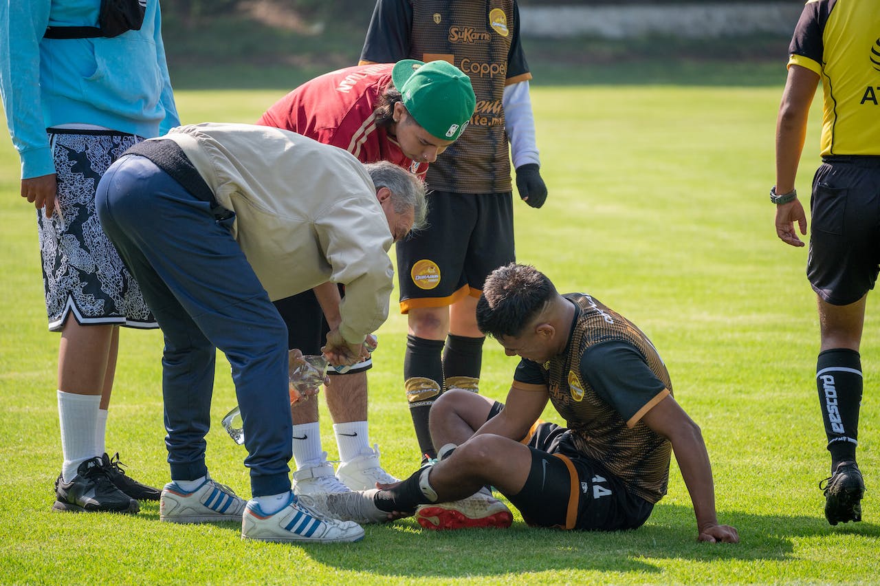
Early female athlete ACL assessment should still start with mechanism, swelling pattern, instability history, and sport context. Non-contact deceleration, cutting, or landing with rapid effusion and a pop remains a classic presentation, but the examination should avoid tunnel vision. Meniscal injury, osteochondral damage, collateral injury, and posterolateral involvement can all change management.
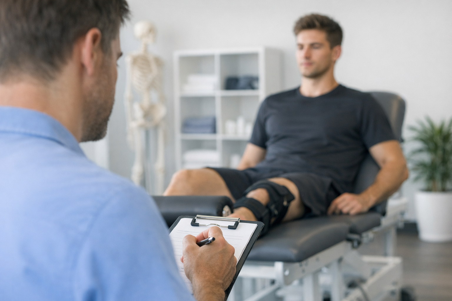
2.1 Clinical examination
The core bedside examination remains highly relevant in ACL injury in female athletes. In the acute or subacute phase, useful maneuvers include Lachman, anterior drawer, pivot shift when feasible, range of motion, effusion grading, and collateral testing.
The Lachman test is often the most practical first-line maneuver, especially when pain or guarding limits more provocative testing. Still, interpretation should be cautious in highly flexible athletes, because baseline laxity may blur the line between physiologic mobility and pathologic asymmetry.
A helpful clinical question is not simply, “Is the knee loose?” but, “Is it looser than the contralateral side in a way that fits the history and symptoms?” That is where understanding knee laxity becomes important.
2.2 Imaging and its limits
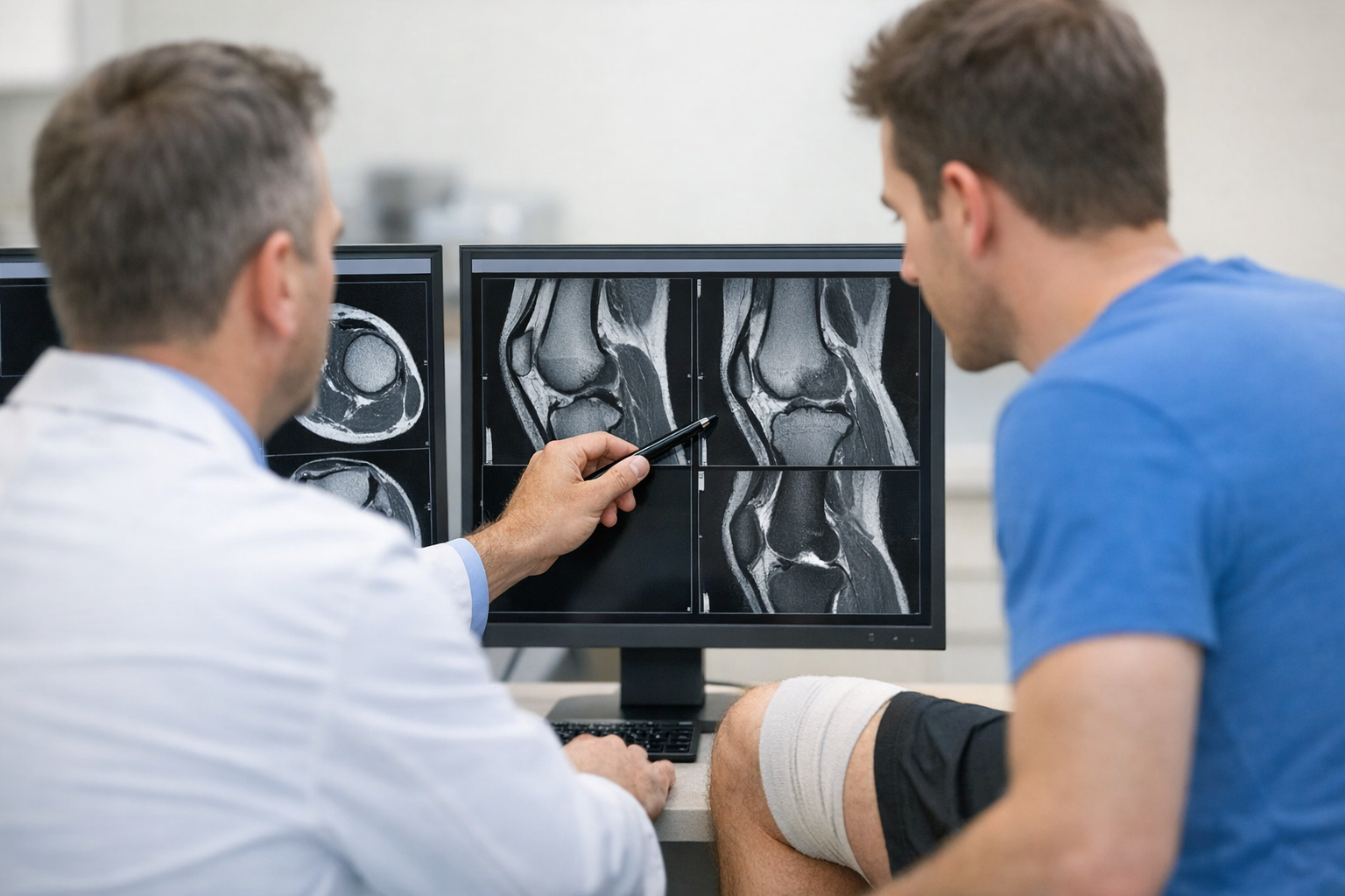
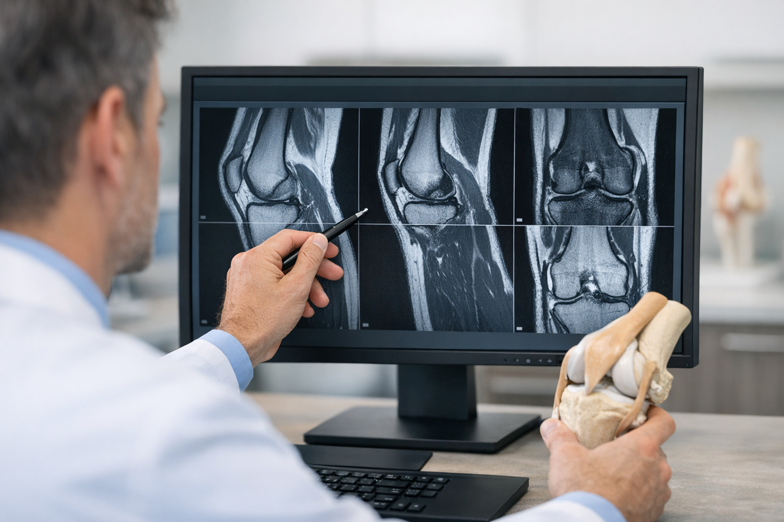
MRI remains important in ACL injury in female athletes, particularly to confirm structural injury and assess meniscus, cartilage, bone bruise pattern, and associated ligament damage. It should not be displaced by any single office tool. At the same time, MRI is not a direct measure of real-time mechanical instability.
In suspected partial tears or equivocal imaging, objective laxity testing may help quantify functional instability that static imaging may not fully capture, but MRI remains complementary and is typically needed for associated injuries and pre-operative planning when reconstruction is considered. This is especially relevant in borderline cases where clinicians are weighing instability symptoms against imaging findings, as discussed in MRI vs arthrometer pathways.
3. Ligament assessment and knee laxity testing ACL pathways
For many clinicians, the next step after history, exam, and MRI review is deciding whether more objective quantification will change management. In ACL injury in female athletes, that question often arises when there is generalized hypermobility, a partial injury pattern, recurrent giving way, or difficulty reconciling symptoms with standard strength milestones.
knee laxity testing ACL pathways are most useful when they quantify side-to-side difference in a reproducible way and when results can be interpreted alongside clinical examination, not in isolation. A practical overview of knee laxity testing can help clinicians choose where such data fit in their workflow.
One relevant concern in female populations is hypermobility. Lindskog et al. (2026) reported an increased hazard ratio of second ACL injury after return to sport for each positive hypermobility test on the Beighton score. The study does not imply that hypermobility alone determines outcome, but it supports closer scrutiny of baseline laxity and reinjury counseling in athletes with multiple positive Beighton items.
This has two practical implications for ACL injury monitoring:
- Baseline generalized laxity should be documented rather than assumed.
- Follow-up should emphasize change over time, not just absolute mobility.
Where objective measurement is clinically relevant, instrumented or robotic arthrometers may complement examination by quantifying anterior translation and side-to-side asymmetry under standardized loading. In some practices, this may be done with a robotic anterior laxity assessment workflow or with a dynamic knee arthrometer approach, particularly when serial measurement is desired.
That said, a single translation value should not be overinterpreted. Pain, guarding, swelling, hamstring activation, and operator workflow all affect data quality. The goal is not to replace judgment, but to strengthen it with repeatable metrics.
4. Rotational knee instability and the pivot shift question
rotational knee instability is often the missing piece in ACL injury in female athletes. Many athletes do not primarily complain of straight-line instability. Instead, they describe mistrust during cutting, turning, or reactive tasks. This is why the pivot shift remains clinically important even though it can be difficult to perform consistently in awake patients.
The challenge is that anterior translation and rotational control are related but not identical. A knee may show moderate anterior laxity yet substantial rotational symptoms, especially if there is associated anterolateral, meniscal, or capsular involvement. Clinicians who want to move beyond static translation alone may find value in dynamic multi-axis workflows when rotational behavior is central to the case.
In higher-grade cases, a structured approach to the pivot shift can clarify whether rotational instability should affect surgical discussion, rehab emphasis, or return-to-sport timing.
Decision aid: when to suspect rotational instability is underappreciated
- Giving-way episodes occur mainly during cutting or pivoting
- Straight-line hop or strength testing looks acceptable, but confidence is low
- Symptoms persist despite apparently good quadriceps recovery
- There is concern for combined injury patterns or residual instability after reconstruction
For ACL injury in female athletes, this matters because return to high-level multidirectional sport depends on more than sagittal-plane control.
5. Monitoring after injury or reconstruction: what recent evidence adds
ACL injury monitoring should be longitudinal, not event-based. A single “clearance day” snapshot rarely captures the full picture in ACL injury in female athletes. Instead, clinicians should track symptoms, swelling response, strength, performance, movement quality, exposure progression, psychological readiness, and stability measures where available.
Collins et al. (2026) reported that females met return-to-sport criteria at lower rates than males 9 months after ACL reconstruction. Without overextending that finding, it supports a cautious interpretation of time-based milestones. Calendar time is not the same as readiness.
This is directly relevant to return to sport after ACL injury. Athletes may look “on schedule” from a postoperative timeline standpoint while still falling short on neuromuscular, psychological, or stability criteria. A more structured framework for return to sport can help avoid overly simplistic decisions.
Similarly, rehabilitation progress should not be inferred from strength alone. The relationship between functional milestones and mechanical status is discussed well in rehab milestones, and it is especially relevant in ACL injury in female athletes where high-demand cutting sports can expose residual deficits.
5.1 Vision, sensorimotor control, and movement adaptation
Yu et al. (2026) explored the effect of visual disruption on lower-limb kinematic and kinetic characteristics in athletes after ACL reconstruction. For clinicians, the practical lesson is not that visual manipulation should become routine for everyone, but that sensory conditions can reveal hidden control deficits. An athlete who performs well in predictable conditions may still struggle when visual information is reduced or decision-making demands rise.
This is one reason ACL injury monitoring benefits from progressive task complexity. Straight-line tests are useful, but they do not replicate reactive sport.
5.2 Psychological readiness and prehabilitation
Fear of reinjury often shapes movement as much as strength does. Ho et al. (2026) found that prehabilitation for patients undergoing ACL reconstruction reduced kinesiophobia levels at 1 year postoperation. For ACL injury in female athletes, that supports addressing confidence and education early, not only late in rehab.
Psychological readiness should therefore be monitored alongside symptoms and function, particularly when athletes appear physically recovered but remain hesitant in deceleration or contact scenarios.
6. Emerging tools, practical interpretation, and next-step decisions
Newer diagnostic approaches may eventually expand how clinicians evaluate ACL injury in female athletes. Allott et al. (2026) examined wearable sensor technology and machine learning for ACL injury diagnosis in an exploratory feasibility study. At present, such tools are promising but should be viewed as early-stage adjuncts rather than definitive clinical solutions.
In day-to-day practice, the most useful monitoring strategy is usually the one that is repeatable, clinically interpretable, and tied to decisions. That may include physical examination, side-to-side strength data, functional testing, patient-reported outcomes, and, when relevant, objective laxity metrics.
For ACL injury in female athletes, a practical clinician-led sequence can look like this:
- Confirm the likely injury pattern with history, examination, and MRI when indicated.
- Document baseline laxity, hypermobility features, effusion, range, and symptom triggers.
- Track progression with repeated clinical review rather than relying on time alone.
- If instability remains unclear, consider objective ACL assessment to quantify side-to-side difference and support interpretation.
- Before clearance, evaluate not just strength but confidence, task complexity, and sport exposure tolerance.
The key pitfall is assuming that asymptomatic straight-line performance equals full readiness. In ACL injury in female athletes, the last deficits to normalize may be rotational control, reactivity, and confidence under pressure.
7. Key takeaways and next steps
ACL injury in female athletes should be assessed through a combined structural, mechanical, and functional lens. Clinical examination remains central. MRI remains complementary and essential for associated injury assessment and planning. Where uncertainty persists, objective ACL assessment may help quantify instability and improve follow-up, especially in athletes with hypermobility, suspected partial injury, or discordant symptoms.
For clinicians, the most important next step is to build monitoring systems that are consistent over time. In ACL injury in female athletes, better decisions often come from repeated integrated assessment, not from any single test. That approach is particularly relevant when planning return to sport after ACL injury, where lower return-to-criteria rates, hypermobility-related reinjury concerns, and residual rotational knee instability may all shape the final decision.
Used carefully, serial examination, targeted imaging, and appropriately selected stability testing can help make ACL injury in female athletes assessment more precise, more individualized, and more clinically responsible.
Clinical references (PubMed)
1) 2026 – Collins et al. – Females Meet Return to Sport Criteria at Lower Rates Than Males 9 Months After ACL Reconstruction. – Orthop J Sports Med – DOI: 10.1177/2325967126S00106 – PMID: 42333090 – PubMed
2) 2026 – Lindskog et al. – Increased Hazard Ratio of a Second ACL Injury After Return to Sport for Each Positive Hypermobility Test on the Beighton Score: A Registry Study. – Sports Med Open – DOI: 10.1186/s40798-026-01054-5 – PMID: 42334734 – PubMed
3) 2026 – Allott et al. – Wearable sensor technology and machine learning for anterior cruciate ligament injury diagnosis: an exploratory feasibility study. – BMJ Open – DOI: 10.1136/bmjopen-2025-107409 – PMID: 42315273 – PubMed
4) 2026 – Yu et al. – Effect of visual disruption on the lower limb kinematic and kinetic characteristics in athletes after anterior cruciate ligament reconstruction. – Front Bioeng Biotechnol – DOI: 10.3389/fbioe.2026.1767092 – PMID: 42339465 – PubMed
5) 2026 – Ho et al. – Prehabilitation for Patients Undergoing Anterior Cruciate Ligament Reconstruction Reduces Kinesiophobia Levels at 1-Year Postoperation. – Sports Health – DOI: 10.1177/19417381261453652 – PMID: 42339880 – PubMed