With the 2026 global football tournament underway across the USA, Mexico, and Canada, clinicians are again seeing intense interest in ACL injury in soccer and how to assess it quickly, accurately, and responsibly. Although this article is not affiliated with that event, the tournament context is a useful reminder that football exposes the knee to high-speed cutting, deceleration, contact, and rotational loads. For sports physicians, orthopaedic surgeons, physiotherapists, and emergency teams, a good soccer ACL injury context starts with mechanism, acute examination, and recognition of combined injury patterns. A structured football ACL injury assessment can improve early triage, imaging decisions, and follow-up planning.
1. Why ACL injury in soccer follows recognisable patterns
ACL injury in soccer is rarely a random event. In many cases, the mechanism points toward the likely pattern of associated damage. Noncontact episodes often involve deceleration, change of direction, landing, or a valgus-rotation collapse. Contact injuries may add direct force, hyperextension, or varus-valgus loading, which can broaden the differential beyond isolated ACL rupture.
This matters because soccer knee ligament injury patterns often include meniscal, chondral, bone bruise, and capsuloligamentous involvement. In current football practice, mechanism-informed assessment helps avoid anchoring too early on a single structure.
Kamada et al. (2026) reported distinct meniscus tear and bone bruise patterns in soccer players with ACL injuries when comparing contact and noncontact mechanisms. Clinically, that supports a more targeted reading of the initial story, especially when MRI is delayed or the acute examination is limited by pain and effusion. If the player describes a pivoting noncontact event with immediate instability, the probability of a true acute ACL tear diagnosis rises, but clinicians should still assess for lateral meniscal injury, osteochondral injury, and peripheral stabiliser involvement.
When pain, swelling, and inability to continue are present, early pathway thinking is useful. A practical triage algorithm can help identify which athletes need urgent imaging, aspiration consideration, protected weight bearing, or specialist review.
2. ACL injury in soccer: first-contact clinical workflow
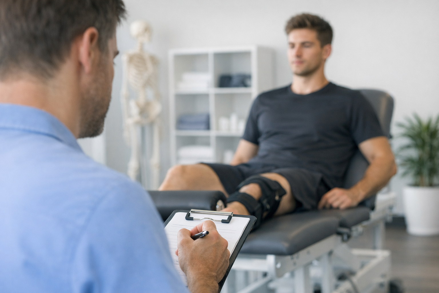
The best early approach to ACL injury in soccer is not one test. It is a sequence. History, inspection, effusion, range, protective spasm, and focused stability testing usually provide more value than any isolated maneuver.
2.1 Key history points
For ACL injury in soccer, ask:
- Was the mechanism noncontact pivot, deceleration, landing, hyperextension, or direct contact?
- Did the athlete feel a pop, shift, or buckle?
- Could they continue playing?
- Was swelling immediate, early, or delayed?
- Is there locking, catching, or inability to fully extend?
- Any prior ACL reconstruction, instability, or contralateral injury?
These details shape the probability of isolated ACL deficiency versus a more complex football knee injury.
2.2 Early examination priorities
In the first 24 to 72 hours, ACL injury clinical workflow should prioritise:
- Effusion and haemarthrosis
- Active and passive extension deficit
- Joint line tenderness
- Collateral tenderness or gapping
- Neurovascular status when trauma is significant
- Ability to weight bear and gait quality
Acutely, exam quality may be reduced by guarding. That is one reason repeat assessment remains important. If the first visit is inconclusive, a structured re-check after swelling and spasm settle may be more diagnostic than forcing a painful exam.
For clinicians refining test choice, these ACL tests are best understood as complementary rather than competitive.
3. Lachman, pivot shift, and rotational clues that change decisions
In suspected ACL injury in soccer, the core bedside tests remain highly relevant, particularly when interpreted in context rather than as binary yes-or-no signs. The usual exam sequence includes Lachman, anterior drawer where appropriate, and assessment for pivot shift and Lachman test findings.
3.1 Lachman first
The Lachman test is often the most practical frontline maneuver in an acute ACL tear diagnosis, especially when compared with tests that need greater knee flexion or less guarding. It helps estimate anterior tibial translation and end-point quality. A soft or absent end point can be more informative than translation alone.
Subtle technical details matter. Patient relaxation, femoral stabilisation, and tibial hand position influence the result. This detailed Lachman guide is useful when acute pain or larger thighs make positioning difficult.
3.2 Pivot shift and rotational instability
Rotational knee instability assessment becomes especially relevant in football because many athletes do not only report forward looseness. They describe a giving-way episode during cut, turn, or landing. That pattern may reflect combined translation and rotation.
The pivot shift can reveal clinically meaningful rotational instability, but it has limitations. In the acutely injured player, pain and guarding can suppress the phenomenon. Under anaesthesia, it may become more apparent. A negative pivot shift in clinic does not exclude functionally important rotational laxity.
This is where mechanism and associated structures matter. Suspicion should increase when there is:
- Lateral-sided pain or bruising
- High-grade giving way during cutting
- Hyperextension pattern
- Concern for anterolateral, meniscal, or posterolateral contribution
For broader context on combining manual findings with structured documentation, see this framework for objective examination.
In professional soccer players after reconstruction, Ackermann et al. (2026) reported that static tibiofemoral rotation remained stable and was associated with posterior tibial slope. While this does not replace bedside assessment, it reinforces that rotational questions in football are not just subjective complaints. They may reflect underlying morphology and stability behaviour that deserve careful interpretation.
4. MRI, repeat examination, and when objective laxity data may help
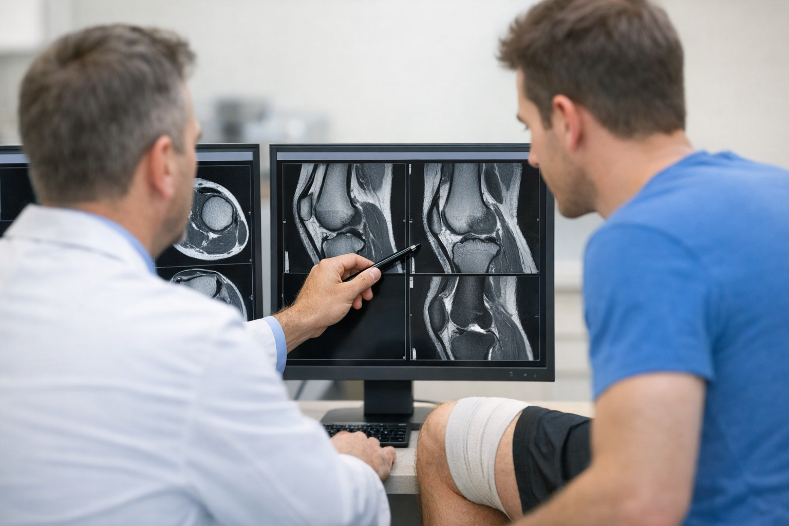
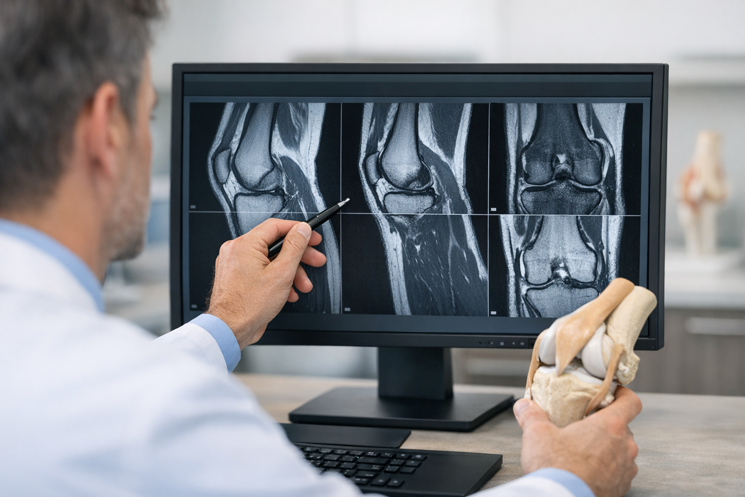
For ACL injury in soccer, MRI remains important, especially when clinicians need to assess meniscus, cartilage, bone bruising, peripheral structures, or plan surgery. However, MRI and examination answer different questions. MRI shows structure. Clinical testing addresses function. Neither should be treated as a complete substitute for the other.
In straightforward cases, concordance between history, Lachman, pivot-related findings, and MRI is common. The challenge is the borderline group: partial injury, equivocal imaging language, guarding that limits exam quality, or persistent symptoms despite a “low-grade” report. In those situations, an MRI vs arthrometer discussion is best framed carefully: objective laxity testing may complement MRI by quantifying side-to-side instability and adding functional information, but MRI is still typically needed to assess associated injuries and for pre-operative planning when reconstruction is considered.
That is particularly relevant in suspected partial tears or a borderline MRI workflow, where the practical question is not only “is the ligament abnormal?” but also “is the knee functionally unstable?”
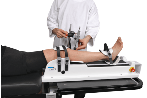
When the clinic pathway includes instrumented assessment, dynamic or robotic measures may help quantify anterior and, in some systems, multi-axis instability in a more reproducible way. A GNRB arthrometer or Dyneelax knee arthrometer may be useful as complementary tools alongside examination and MRI, especially when documentation of side-to-side difference or dynamic response could clarify a difficult football knee.
For clinicians interested in broader implementation, this multi-axis workflow shows how objective data can be integrated without displacing clinical judgment.
If MRI confirms ACL deficiency with meniscal involvement, the pattern may influence instability interpretation. This becomes particularly relevant in combined lesions such as the ACL-deficient knee with lateral meniscal tears, where this report on lateral meniscal tears is clinically relevant to the football setting.
5. Decision aid for suspected ACL injury in soccer
A simple decision framework can make ACL injury in soccer assessment more consistent across the emergency department, sports clinic, and orthopaedic review.
5.1 Practical 5-step decision aid
- Confirm mechanism – Noncontact pivot, deceleration, or landing raises suspicion; contact mechanism broadens the ligament differential.
- Assess the swollen knee – Early haemarthrosis, loss of extension, and instability symptoms support significant internal derangement.
- Perform focused tests – Prioritise Lachman, then add pivot-related and collateral assessment as tolerated.
- Look for combined injury clues – Joint line pain, locking, hyperextension pattern, or lateral/posterolateral findings may change urgency and management.
- Decide on next step – MRI, repeat exam, protected rehabilitation, urgent orthopaedic input, or complementary objective laxity testing depending on the case.
This type of football ACL injury assessment is especially important when acute pain masks instability. If the athlete cannot relax, the clinician should not overstate certainty. Instead, document what is known, what remains uncertain, and why follow-up testing or imaging is needed.
In short, ACL injury in soccer should be approached as a clinician-led decision process, not a one-visit verdict.
6. Return-to-play thinking starts at diagnosis
Although definitive return planning comes later, the first assessment of ACL injury in soccer should already consider likely sport demands. Soccer places repeated cutting, perturbation, deceleration, and jump-landing loads on the knee. That means the diagnosis phase should document not just pain and swelling, but also instability phenotype, associated pathology, and baseline performance deficits.
This is where the language of return to play ACL football begins. Not because the player is close to returning, but because early decisions influence later readiness criteria, surgery choices, rehabilitation sequencing, and expectations.
Andrić et al. (2026) examined squat jump and bilateral and unilateral countermovement jump performance in soccer players at 6 and 9 months after ACL reconstruction, underscoring that performance recovery is not captured by time alone. Similarly, Rodríguez et al. (2026) described periodization of physical exercise in rehabilitation after ACL reconstruction in a professional soccer player, which aligns with the practical need for staged, criteria-informed progression rather than calendar-based clearance.
Even surgical adjuncts aimed at rotational control should be interpreted carefully. In professional soccer players, Ackermann et al. (2026) reported that lateral extra-articular tenodesis did not affect tibiofemoral axial rotation in primary ACL reconstruction. For clinicians, that is a reminder that managing rotational knee instability assessment and functional sport stability involves more than one operative or imaging variable.
Once treatment is underway, objective reassessment becomes important. These return-to-sport criteria can help frame follow-up around stability, function, and progression rather than symptoms alone.
7. Key takeaways and next steps
ACL injury in soccer is common, but the most useful assessments are still the ones that combine mechanism, repeatable examination, and context-aware imaging. In football, the main pitfall is to think only in terms of a torn ACL or not torn ACL. Real decisions usually depend on whether there is functional instability, rotational involvement, meniscal injury, extension block, or combined ligament damage.
For day-one care, remember these points:
- ACL injury in soccer often presents with recognisable noncontact or contact patterns.
- Early Lachman findings may be more practical than pivot shift in a guarded acute knee.
- MRI remains complementary and important for associated injuries and planning.
- Objective knee laxity testing may help in selected equivocal or partial-tear pathways, but it should complement, not replace, MRI and examination.
- ACL injury in soccer management should connect diagnosis to later return-to-play demands from the start.
The next step is usually one of three things: urgent specialist referral when the pattern is complex, MRI when structural definition is needed, or repeat targeted review when acute pain limits exam confidence. In all cases, clinician judgment remains central.
Clinical references (PubMed)
1) 2026 – Kamada et al. – Distinct meniscus tears and bone bruise patterns in soccer players with anterior cruciate ligament injuries: A comparative study of noncontact and contact mechanisms. – Knee – DOI: 10.1016/j.knee.2026.104458 – PMID: 42000385 – PubMed
2) 2026 – Andrić et al. – Squat Jump and Bilateral and Unilateral Countermovement Jump Performance in Soccer Players 6 and 9 Months After Anterior Cruciate Ligament Reconstruction. – Medicina (Kaunas) – DOI: 10.3390/medicina62050807 – PMID: 42195060 – PubMed
3) 2026 – Ackermann et al. – Static tibiofemoral rotation remains stable after ACL reconstruction and is associated with posterior tibial slope in professional soccer players. – Knee Surg Sports Traumatol Arthrosc – DOI: 10.1002/ksa.70362 – PMID: 41758997 – PubMed
4) 2026 – Ackermann et al. – Lateral extra-articular tenodesis does not affect tibiofemoral axial rotation in primary ACL reconstruction in professional soccer players. – Knee Surg Sports Traumatol Arthrosc – DOI: 10.1002/ksa.70283 – PMID: 41566872 – PubMed
5) 2026 – Rodríguez et al. – Periodization of physical exercise in the rehabilitation of a professional soccer player following anterior cruciate ligament reconstruction: A case report. – J Bodyw Mov Ther – DOI: 10.1016/j.jbmt.2025.12.006 – PMID: 41927228 – PubMed