
In some ACL-injured knees, the MRI appearance and the clinical picture do not match. A patient may have a subtle or borderline scan, yet demonstrate a clear pivot shift test, recurrent giving-way, or high-demand functional failure. That gap matters because rotational knee instability is a dynamic problem, not only a structural imaging finding. For orthopaedic surgeons, sports physicians, radiologists, and physiotherapists, the key question is not whether MRI is useful, but when MRI may under-represent the real-world severity of instability and what additional assessment helps clarify risk, treatment planning, and return-to-sport decisions.
1. Why rotational knee instability can look mild on MRI but feel severe clinically
Rotational knee instability is often most apparent during movement, load acceptance, cutting, deceleration, or combined valgus-internal rotation stress. MRI, by contrast, is a static study acquired in a non-weight-bearing setting. That mismatch is one reason an ACL injury that appears partial, low grade, or difficult to classify on MRI may still produce major functional knee instability.
This is especially relevant in anterolateral rotatory instability, where the ACL may not be the only structure involved. Injury patterns involving the anterolateral complex, capsular structures, meniscal restraint, bony morphology, or occult secondary stabilizers can amplify pivoting symptoms without always producing a dramatic MRI report.
Clinically, the patient history may be more revealing than the scan wording. Red flags include recurrent pivoting episodes, apprehension with change of direction, inability to trust the knee despite near-normal straight-line function, and persistent instability after ACL reconstruction suggesting ACL residual instability.
For clinicians working through this discrepancy, an objective exam is often the best starting point. It helps place imaging findings in the context of symptoms, mechanism, and side-to-side function.
1.1 Common reasons MRI under-represents functional severity
- Static imaging cannot reproduce the pivoting mechanism
- Partial tears may preserve visual continuity despite reduced restraint
- Associated anterolateral or capsular injury may be subtle
- Bone bruise, notch sign, or fracture patterns may be underweighted in reporting
- Residual graft laxity or altered stiffness may be clinically important before gross MRI abnormality is obvious
2. Clinical clues that matter more than MRI wording in rotational knee instability
When evaluating rotational knee instability, the exam should focus on whether the knee subluxes dynamically, not only whether the ACL fibers are seen. The pivot shift test remains central because it reflects the combined loss of restraint that patients often perceive as instability in daily sport-specific function.
A high-grade pivot shift has practical implications even when MRI findings seem modest. In fact, our related discussion of pivot shift interpretation is useful when deciding whether apparent imaging severity is actually underestimating the problem.
Nakamae et al. (2026) examined factors affecting quantitative pivot shift values in ACL-injured knees using a navigation system. For clinicians, that reinforces an important principle: pivot shift severity is not a simple yes-or-no phenomenon. It is influenced by multiple anatomical and biomechanical factors, which helps explain why two knees with similar MRI reports may behave very differently in practice.
History and examination findings that should raise concern include:
- Reproducible giving-way during cutting or pivoting
- Apprehension on rotational loading despite acceptable sagittal laxity
- A clearly asymmetric pivot shift compared with the contralateral side
- Post-reconstruction symptoms despite apparently intact graft morphology
- Mismatch between rehab progress and instability complaints
In these cases, rotational knee instability should not be downgraded simply because the report says partial tear, sprain, graft continuity preserved, or no major secondary lesion.
3. MRI signs that may hint at underestimated rotational knee instability
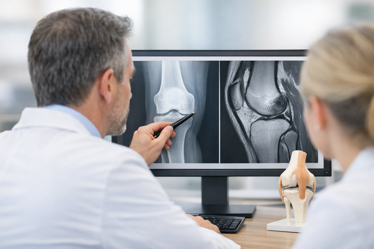
Although MRI may under-represent dynamic severity, it still provides essential structural information. It remains complementary, particularly for meniscal, chondral, osseous, and pre-operative assessment. The issue is not that MRI is unhelpful, but that MRI findings should be interpreted as part of a broader instability model.
Several imaging clues may increase suspicion that rotational knee instability is more clinically important than the headline MRI impression suggests.
3.1 Lateral compartment and anterolateral clues
Dimitriou et al. (2021) reported that the deep lateral femoral notch sign can be a reliable diagnostic clue for combined ACL and anterolateral ligament injury. In practical terms, that means a radiographic or MRI-associated osseous clue may point toward more meaningful anterolateral rotatory instability than a simple ACL descriptor alone implies.
Willinger et al. (2026) found an association between posterolateral tibial plateau fractures and anterolateral complex injuries in primary ACL injury. For radiologists and surgeons, this matters because subtle lateral-sided bony injury may act as a marker of a broader rotational injury pattern, even if the principal MRI conclusion appears relatively limited.
3.2 Post-operative MRI can also mislead
After reconstruction, MRI continuity or ligamentization appearance does not always guarantee normal function. Kawoosa et al. (2026) evaluated early clinical and MRI assessment of graft ligamentization, illustrating the broader point that graft appearance and clinical behavior are related but not interchangeable.
Similarly, Yeganeh et al. (2025) examined the impact of ligamentous adhesion to the posterior cruciate ligament on radiological, arthroscopic, and clinical outcomes after ACL reconstruction. This is a useful reminder that postoperative imaging findings and postoperative symptoms may diverge, and that persistent ACL residual instability may require functional assessment rather than image interpretation alone.
Where MRI findings are equivocal, clinicians may also find value in reviewing a structured MRI vs arthrometer comparison, especially in cases where instability symptoms are out of proportion to scan wording.
4. Decision points: when to escalate assessment beyond MRI
Not every patient with an ACL injury and mild MRI findings needs expanded testing. But when rotational knee instability remains a real clinical concern, relying on MRI alone may leave important uncertainty unresolved.
Consider escalation when one or more of the following are present:
- Borderline or partial ACL report with clear instability symptoms
- Positive or high-grade pivot shift out of proportion to MRI language
- Suspected combined injury pattern involving lateral or capsular structures
- Failure to progress despite adequate rehabilitation
- Persistent instability after ACL reconstruction
- High-demand athletes where treatment decisions depend on functional severity
4.1 A short decision aid
- Start with mechanism, symptoms, and standard clinical examination.
- Review MRI for ACL morphology, bone bruising, notch sign, lateral-sided clues, meniscus, and cartilage.
- If the exam and MRI align, proceed with the usual management pathway.
- If they do not align, quantify dynamic knee laxity and side-to-side asymmetry.
- Use the combined picture to guide nonoperative planning, surgical indication, graft strategy, or return-to-sport timing.
This is especially helpful in borderline cases, where MRI remains complementary and often still needed for associated injury assessment and pre-operative planning, but does not fully answer the question of functional instability.
Clinicians also benefit from understanding the broader menu of diagnostic tests rather than treating MRI as the single decision-maker.
5. Where objective laxity testing fits in rotational knee instability
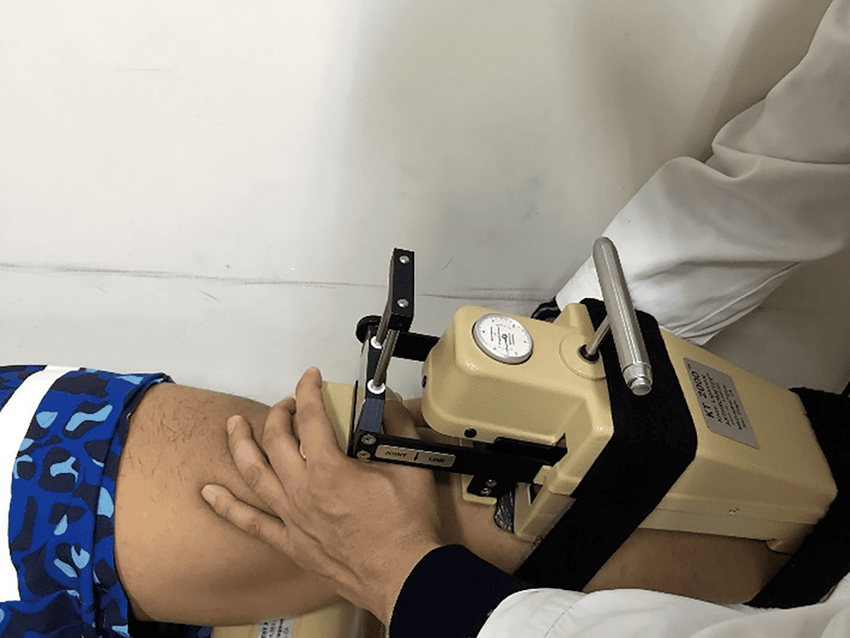
When rotational knee instability is suspected but MRI under-represents functional severity, objective laxity testing can add the missing dynamic layer. It does not replace MRI. Instead, it complements MRI and clinical examination by quantifying side-to-side instability, mechanical response, and in some pathways may help clarify equivocal presentations or triage who needs further imaging review and specialist referral.
This is where instrumented knee laxity testing and broader objective rotational laxity assessment become clinically relevant. Depending on the workflow, instrumented assessment may help characterize anterior translation behavior, compliance, stiffness, and multi-axis features that contribute to instability perception, particularly when conventional examination is limited by guarding or when postoperative symptoms are difficult to interpret.
For clinics integrating this approach, laxity testing should be framed as part of a combined assessment model. In more advanced pathways, dynamic knee laxity testing can support interpretation of rotational patterns that are not obvious on static imaging alone.
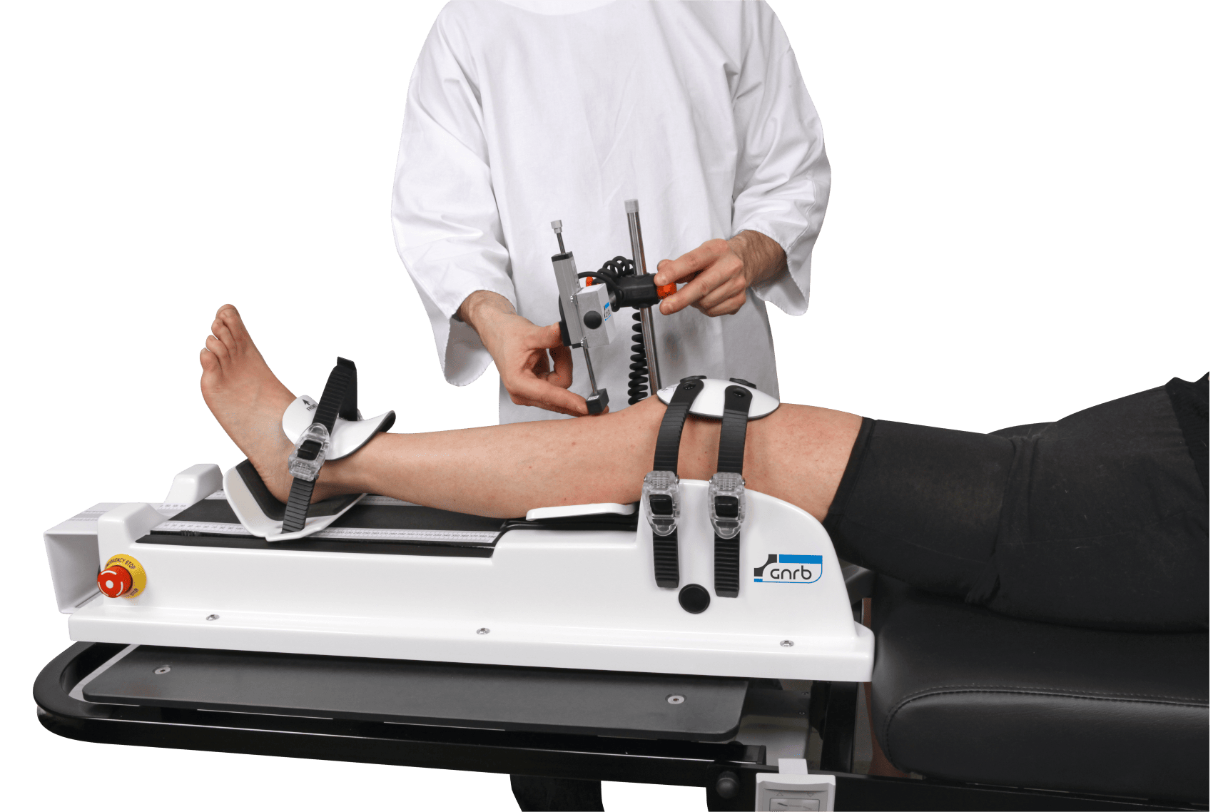
Where device-based assessment is used, clinicians may consider a GNRB arthrometer assessment for objective anterior laxity profiling and a Dyneelax evaluation when a broader robotic or dynamic knee assessment pathway is available.
One practical point is that translation alone may not tell the whole story. Material behavior and functional restraint also matter. That is why some clinicians increasingly review stiffness metrics and compliance patterns when symptoms suggest instability despite less dramatic absolute displacement values.
In selected postoperative or return-to-play cases, this objective layer may be more informative than MRI appearance alone when discussing confidence, progression, and reinjury risk.
6. Interpreting rotational knee instability in rehab, surgery, and return to sport
Rotational knee instability has consequences beyond diagnosis. It affects whether rehabilitation is likely to succeed without recurrent episodes, whether extra attention should be paid to combined injury patterns, and whether return-to-sport decisions are being made on appearance or actual mechanical stability.
For physiotherapists, one of the common pitfalls is assuming that improving strength, hop scores, or symptom tolerance means the instability problem has resolved. In some athletes, performance compensation can temporarily mask persistent functional knee instability.
That is why it helps to align rehabilitation milestones with measured stability. Our summary on rehab progress addresses this directly.
For surgeons, the same mismatch applies after reconstruction. A graft that looks acceptable on MRI may still coexist with clinically meaningful ACL residual instability, especially if there is unresolved rotational laxity, tunnel-related mechanics, associated lateral injury, or altered graft behavior under load.
For sports medicine return-to-play pathways, a normal-appearing scan should not automatically outweigh symptoms, exam findings, or quantified instability. Objective thresholds remain clinician-led and context-specific, but the principle is consistent: return to sport should reflect stability, not imaging reassurance alone. A related framework is available in return-to-sport criteria.
Ultimately, rotational knee instability should be interpreted as a functional diagnosis informed by imaging, not defined by imaging alone.
7. Key takeaways and next steps
Rotational knee instability may be clinically severe even when MRI appears modest. This is most likely when the instability is dynamic, combined, or influenced by anterolateral structures, subtle lateral-sided injury, postoperative mechanics, or altered tissue behavior under load.
The practical response is not to dismiss MRI, but to place it correctly. MRI remains essential for associated injuries and operative planning. However, if symptoms, exam findings, or the pivot shift test suggest more than the scan shows, the next step is a clinician-led synthesis of history, examination, and objective functional assessment.
Next steps for practice:
- Reassess the mechanism and instability history carefully
- Grade the pivot shift and compare sides
- Review MRI for subtle lateral or combined injury markers
- Use objective assessment when MRI and function do not match
- Base treatment and return-to-sport decisions on the whole stability picture
In short, rotational knee instability is easiest to miss when clinicians ask only what the MRI shows, instead of also asking how the knee behaves.
Clinical references (PubMed)
1) 2026 – Willinger et al. – Posterolateral tibial plateau fractures are associated with anterolateral complex injuries of the knee in primary ACL injury. – Knee Surgery, Sports Traumatology, Arthroscopy – DOI: 10.1002/ksa.70393 – PMID: 41979380 – PubMed
2) 2021 – Dimitriou et al. – The deep lateral femoral notch sign: a reliable diagnostic tool in identifying a concomitant anterior cruciate and anterolateral ligament injury. – Knee Surgery, Sports Traumatology, Arthroscopy – DOI: 10.1007/s00167-020-06278-w – PMID: 32974801 – PubMed
3) 2025 – Yeganeh et al. – Impact of Ligamentous Adhesion to the Posterior Cruciate Ligament on Radiological, Arthroscopic, and Clinical Outcomes One Year After ACL Reconstruction: A Cohort Study : – Galen Med J – DOI: 10.31661/gmj.v14i.3589 – PMID: 42038857 – PubMed
4) 2026 – Nakamae et al. – Factors Affecting the Quantitative Value of the Pivot Shift Test Using a Navigation System in Anterior Cruciate Ligament Injured Knees. – J Knee Surg – DOI: 10.1055/a-2865-3249 – PMID: 42057414 – PubMed
5) 2026 – Kawoosa et al. – Anterior Cruciate Ligament Reconstruction Using Tibial Attachment – Preserving Hamstring Grafts: Early Clinical and Magnetic Resonance Imaging Assessment of Graft Ligamentization. – J Orthop Case Rep – DOI: 10.13107/jocr.2026.v16.i05.7348 – PMID: 42130994 – PubMed