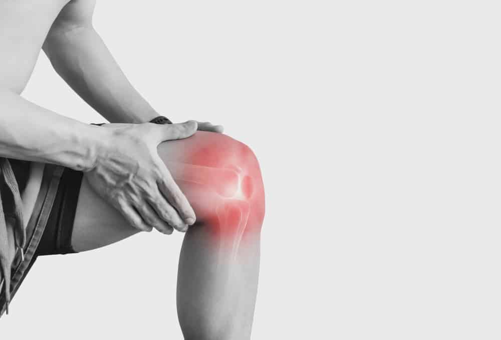

Objective knee examination has become increasingly important in modern ligament assessment because it helps translate a subjective impression of instability into measurable findings. For orthopaedic surgeons, sports physicians, physiotherapists, and researchers, this matters most when manual knee ligament tests are limited by guarding, swelling, examiner experience, or borderline injury patterns. In practice, a structured approach combines history, manual examination, imaging, and instrumented knee laxity testing to clarify how much instability is present, in which direction, and whether it is functionally relevant. Rather than replacing MRI, an objective knee examination adds quantified information that may improve clinical reasoning in ACL and rotational instability workups.
1. Why objective knee examination matters in ligament assessment
The classic knee exam remains indispensable, but it has known variability. A Lachman test can feel clearly abnormal in one patient and equivocal in another, particularly in acute injury, muscular guarding, or partial tears. This is where quantified knee instability becomes useful: it helps define side-to-side asymmetry, characterize load-response behavior, and track change over time.
For ACL-deficient knees, clinicians are often trying to answer several distinct questions:
- Is there measurable anterior cruciate ligament laxity?
- Is the instability mainly anterior, rotational, or combined?
- How large is the side-to-side difference knee finding compared with the contralateral limb?
- Do imaging findings match the patient’s functional instability?
- Has reconstruction or rehabilitation actually restored stability?
An objective knee examination is especially valuable when these questions do not have the same answer. A knee may look structurally abnormal on MRI but show limited dynamic translation, or the reverse may occur in clinically unstable knees with incomplete or equivocal imaging findings. This is one reason knee laxity measurement has become more relevant in both preoperative and follow-up settings.
Recent imaging-based evidence also supports the concept that tibial position and subluxation relate to instability rather than only morphology. Hamada et al. (2026) examined anterior tibial subluxation on weight-bearing extended lateral radiographs in ACL-deficient knees, highlighting the clinical interest in measurable displacement beyond a purely manual exam. Likewise, Murakami et al. (2026) reported that increased preoperative lateral anterior tibial subluxation on MRI was associated with increased knee laxity after anatomic ACL reconstruction, reinforcing the link between structural alignment features and postoperative instability risk.
2. Manual tests still matter, but they are not the whole story
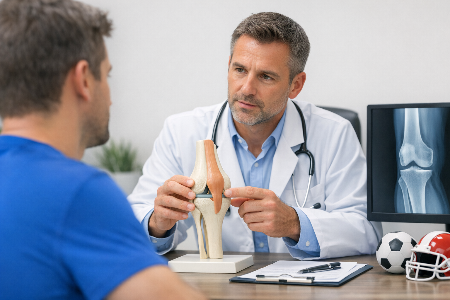
A careful objective knee examination starts with the manual exam, not in competition with it. The clinician still needs to assess swelling, range of motion, effusion, endpoint quality, apprehension, and associated ligament or meniscal injury. Manual testing remains the language of everyday orthopaedic decision-making.
2.1 Key manual tests and what they contribute
The most common manual knee ligament tests in suspected ACL injury include the Lachman test, anterior drawer, pivot shift, and, where relevant, collateral and posterolateral corner testing. The Lachman test remains central because it is often the most practical bedside maneuver for estimating anterior translation and endpoint quality.
Still, manual testing has limitations:
- Examiner dependence affects reproducibility.
- Pain and guarding can mask laxity.
- Large hands vs small knees or vice versa can change the feel of translation.
- Partial ACL tears may produce subtle findings.
- Rotational instability may be underappreciated if only anterior tests are emphasized.
This is why an objective knee examination often works best as a layered process: manual test first, quantified confirmation second, imaging correlation third. A broad overview of this framework is well illustrated in these ACL tests, which help place instrumented and manual methods in the same diagnostic pathway.
2.2 The pivot shift is clinically rich, but hard to standardize
The pivot shift assessment remains one of the most clinically meaningful tests because it reflects dynamic anterolateral rotatory instability rather than isolated anterior translation. A high-grade pivot shift often changes surgical planning, counseling, and concern about residual instability.
At the same time, pivot shift grading can vary with anesthesia status, patient relaxation, and examiner technique. For that reason, objective knee examination is increasingly discussed not just in terms of anterior displacement but also in terms of repeatable rotational or multi-axis instability metrics. The clinical implications of a pronounced pivot shift are discussed in more detail in this guide to pivot shift.
Evidence in this area continues to evolve. Zhang et al. (2026) reviewed risk factors for high-grade pivot shift in ACL tears combined with lateral meniscus posterior root tears, emphasizing that associated lesions can materially influence rotational instability. In a treatment-focused context, Ni et al. (2025) reported that lateral extra-articular tenodesis notably reduced residual instability and anterior tibial translation in patients with ACL injury and a high-grade pivot-shift phenomenon, which further supports careful preoperative and postoperative instability characterization.
3. How to interpret quantified laxity in an objective knee examination
An objective knee examination becomes clinically meaningful when the numbers are interpreted in context rather than treated as isolated outputs. The goal is not simply to record displacement, but to understand what that measurement means for diagnosis, reconstruction planning, rehabilitation, and return to sport.
The most useful principles include:
- Compare sides, not just absolute values.
- Assess whether the instability pattern fits the suspected injured structure.
- Consider timing: acute, chronic, postoperative, or post-reinjury.
- Integrate with symptoms such as giving-way, cutting apprehension, and recurrent effusions.
- Interpret numbers alongside MRI, not instead of MRI.
In ACL pathways, the side-to-side difference knee measurement is often one of the most practical outputs because it adjusts for individual anatomy and baseline laxity. This may help clarify whether apparent translation is clinically relevant or simply reflects generalized looseness. It also makes serial follow-up more useful than a one-time isolated number.
Beyond static displacement, clinicians are increasingly interested in dynamic knee laxity measurement and load-response behavior. Translation under a single force point can be informative, but stiffness, compliance, and response across loading phases may better reflect graft behavior or residual instability patterns. This is why a more advanced objective knee examination may include concepts beyond simple millimeter translation, as outlined in this discussion of why compliance and stiffness can add value.
A practical decision aid:
- Start with history and manual exam findings.
- Confirm whether quantified anterior translation supports suspected ACL insufficiency.
- If rotational symptoms or pivot shift are prominent, look beyond pure sagittal laxity.
- Use MRI to define associated meniscal, chondral, and bony pathology.
- Reassess with the same framework after surgery or during rehabilitation.
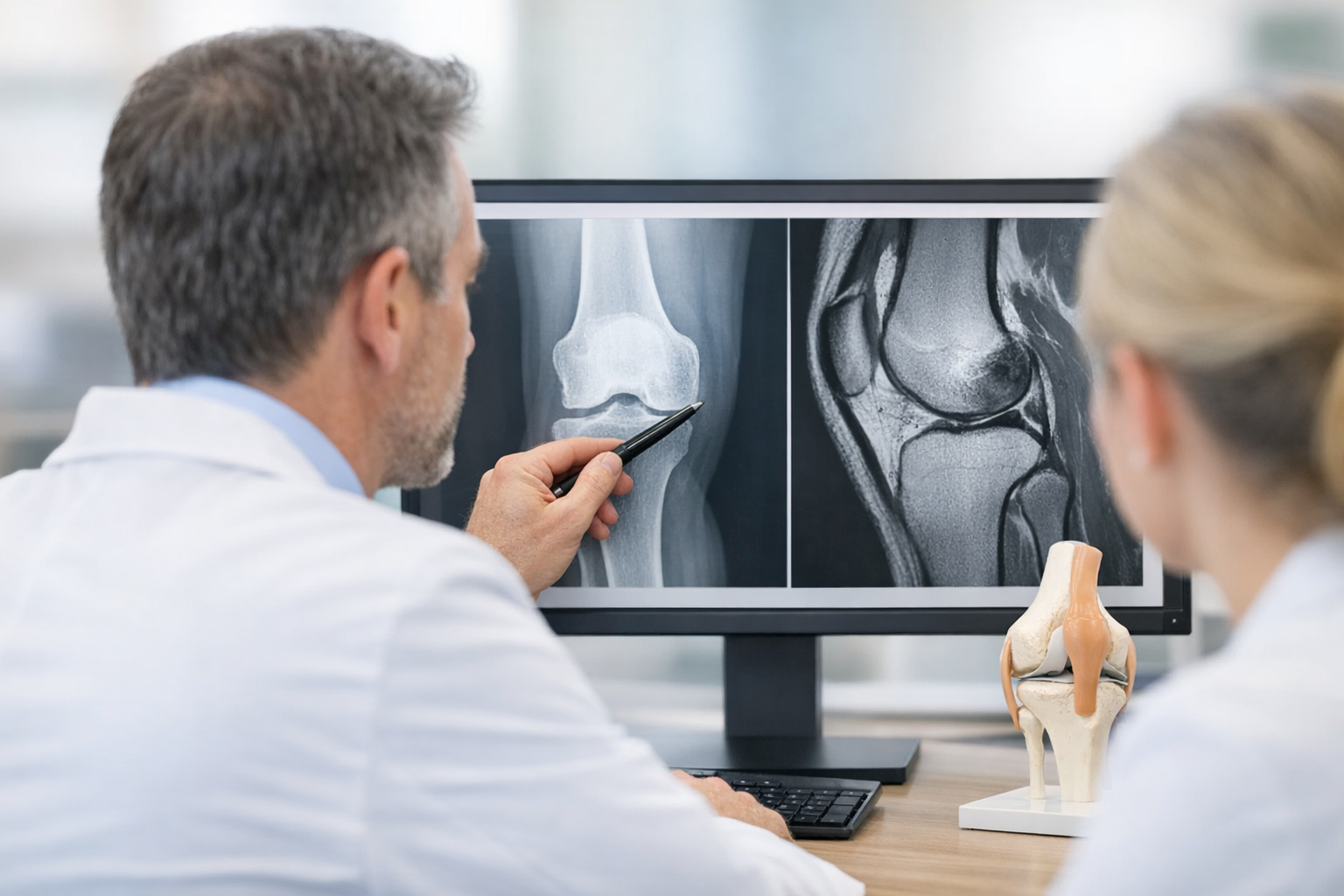
In equivocal or suspected partial ACL tears, objective knee examination may add diagnostic value by quantifying functional instability that MRI does not fully capture. However, MRI remains complementary and is typically needed to assess associated injuries and for preoperative planning when reconstruction is considered. The overlap and distinction between these tools are discussed in this article on MRI vs arthrometer.
4. Where instrumented and robotic assessment fit clinically
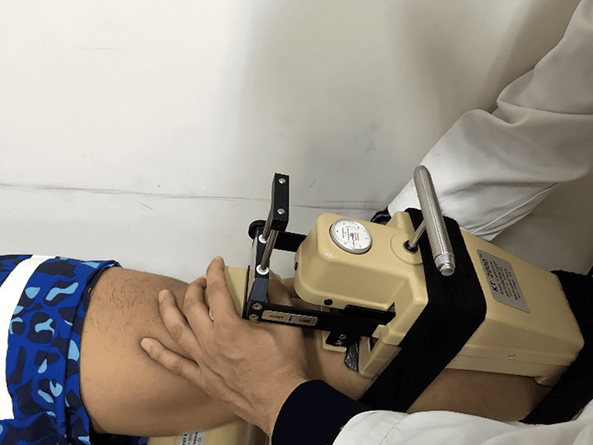
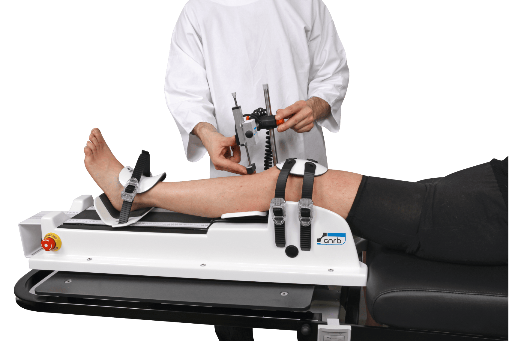
Instrumented knee laxity testing can strengthen an objective knee examination by improving repeatability and reducing reliance on examiner feel alone. In practice, these systems may quantify anterior tibial translation, force-displacement behavior, and in some workflows aspects of dynamic knee laxity measurement that are difficult to capture consistently with manual examination.
This matters most in three situations:
- Borderline or partial ACL presentations where manual tests are subtle.
- Preoperative stratification when the degree of instability may influence planning.
- Postoperative follow-up when clinicians need trendable metrics rather than general impressions.
When clinicians discuss an arthrometer knee evaluation, the key point is not that the device replaces clinical expertise, but that it may support a more reproducible examination pathway. This can be relevant for both standard sagittal assessment and broader multi-axis workflows. Examples of these complementary approaches include the GNRB arthrometer for quantified anterior translation assessment and the Dyneelax system for a broader robotic knee laxity evaluation approach.
A modern objective knee examination may therefore combine manual maneuvers, side-to-side translation values, and dynamic profiling rather than relying on a single test. This is particularly relevant when rotational symptoms are out of proportion to simple anterior laxity, as explored in this multi-axis workflow.
5. ACL laxity and rotational instability: linking numbers to decisions
For ACL-focused practice, the challenge is not merely identifying laxity but understanding which type of instability is driving symptoms and risk. Anterior cruciate ligament laxity alone does not fully describe the unstable knee. Some patients have notable anterior translation with relatively modest pivot phenomena, while others demonstrate substantial rotational instability despite less dramatic sagittal displacement.
This is where an objective knee examination can sharpen treatment thinking. If quantified anterior translation is elevated and the pivot shift assessment is also high grade, clinicians may become more alert to combined pathology such as meniscal root injury, anterolateral complex involvement, or broader rotatory insufficiency.
Zhang et al. (2026) support this perspective by identifying risk factors for high-grade pivot shift in the setting of ACL tears with lateral meniscus posterior root tears. Meanwhile, Ni et al. (2025) suggest that addressing anterolateral rotational instability with lateral extra-articular tenodesis may reduce residual instability in selected high-grade pivot-shift cases. These studies do not create a one-size-fits-all rule, but they do reinforce a principle: objective knee examination should inform phenotype-specific management rather than generic ACL treatment alone.
Computational modeling may also expand future interpretation of injury pattern and instability behavior. Sharabi et al. (2024) explored refined computational modeling of ACL tear injury patterns, which may eventually help connect biomechanical mechanisms, lesion morphology, and observed laxity behavior more precisely.
Clinically, a useful summary is:
- Anterior translation helps define sagittal ACL insufficiency.
- Pivot shift helps capture dynamic rotational abnormality.
- Associated lesions often explain why instability feels worse than one metric suggests.
- Quantified knee instability is most valuable when it changes management or follow-up decisions.
6. Objective knee examination in follow-up, rehab, and return to sport
An objective knee examination is not only a diagnostic tool. It also has value during recovery, especially when symptoms, confidence, and performance do not align neatly. A patient may pass functional drills yet still demonstrate measurable asymmetry, or may report subjective insecurity despite acceptable strength testing.
That is why quantified stability can complement milestone-based rehabilitation. It adds another layer to clinician-led judgment rather than acting as a standalone clearance tool. In practice, this may be particularly useful after ACL reconstruction, revision surgery, or return-to-cutting progression.
For example, return-to-sport decisions are stronger when symptoms, strength, neuromuscular control, exposure tolerance, and objective knee examination findings all point in the same direction. This perspective is developed further in return-to-sport criteria for objective stability.
Similarly, rehabilitation checklists can miss persistent laxity if they focus only on time, swelling, range, and hop performance. Quantified follow-up may reveal residual instability that deserves continued monitoring or further surgeon review. This is one reason clinicians may pair milestone tracking with the concepts discussed in rehab milestones.
Objective knee examination is most helpful in follow-up when the same method is used consistently and interpreted alongside the patient’s function, sport demands, and exam findings.
7. Key takeaways and next steps
Objective knee examination is best understood as a complementary framework, not a single test. It strengthens ligament assessment by combining clinical examination, quantified translation or dynamic response, and imaging correlation. For ACL pathways in particular, it can help define quantified knee instability, clarify side-to-side difference knee findings, and support more consistent follow-up.
The practical next step is simple:
- Use manual examination to identify the likely instability pattern.
- Add quantified assessment when laxity needs clearer definition.
- Use MRI to assess associated injuries and surgical planning needs.
- Reassess stability over time, especially after reconstruction or before return to sport.
For clinicians working with ACL and rotational instability, the most useful objective knee examination is the one that improves decisions without oversimplifying them. Diagnosis and treatment remain clinician-led, but objective data can make that judgment more reproducible, especially when manual findings and symptoms are not perfectly aligned.
Clinical references (PubMed)
1) 2026 – Hamada et al. – Reliability and clinical utility of anterior tibial subluxation on weight-bearing extended lateral knee radiographs in anterior cruciate ligament-deficient knee. – J ISAKOS – DOI: 10.1016/j.jisako.2026.101098 – PMID: 41831688 – PubMed
2) 2026 – Murakami et al. – Increased Preoperative Lateral Anterior Tibial Subluxation by Magnetic Resonance Imaging Is Associated With Increased Knee Laxity After Anatomical Anterior Cruciate Ligament Reconstruction. – Arthroscopy – DOI: 10.1002/arj.70080 – PMID: 41866322 – PubMed
3) 2024 – Sharabi et al. – Predictive Refined Computational Modeling of ACL Tear Injury Patterns. – Bioengineering (Basel) – DOI: 10.3390/bioengineering11050413 – PMID: 38790281 – PubMed
4) 2026 – Zhang et al. – Risk factors for high-grade pivot shift in patients with anterior cruciate ligament tears combined with lateral meniscus posterior root tears: a systematic review and meta-analysis. – Front Med (Lausanne) – DOI: 10.3389/fmed.2026.1813111 – PMID: 42180738 – PubMed
5) 2025 – Ni et al. – Lateral Extra-articular Tenodesis Notably Reduced Residual Knee Instability and Anterior Tibial Translation in Patients With an Anterior Cruciate Ligament Injury Combined With a High-Grade Pivot-Shift Phenomenon. – Orthop J Sports Med – DOI: 10.1177/23259671251323905 – PMID: 40182565 – PubMed