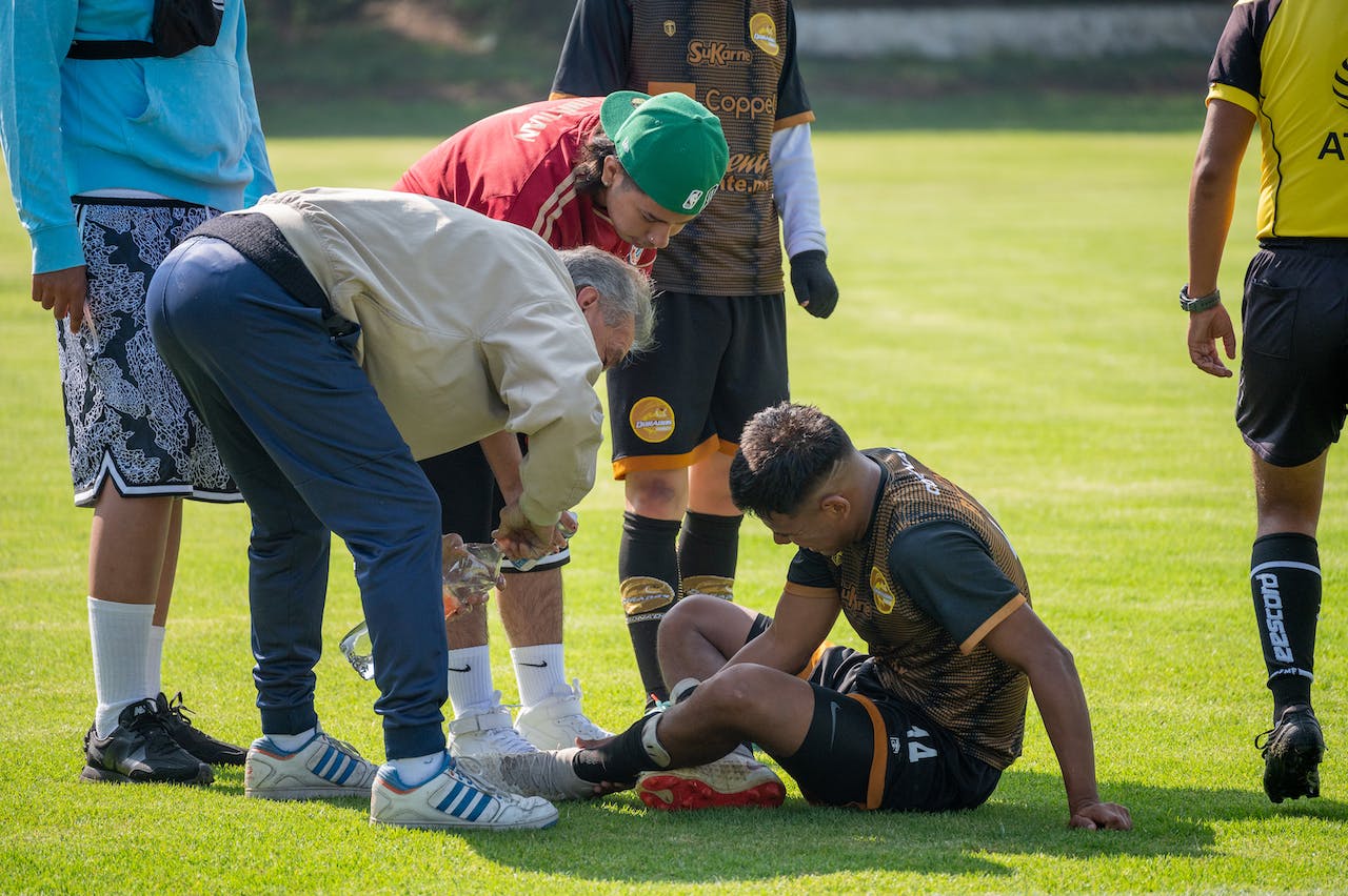
Not every anterior cruciate ligament injury announces itself with a loud “pop.” In real-world practice, ACL tear without pop is a common source of diagnostic delay, especially when swelling is mild, the athlete finishes the session, or the story is vague. This creates the perfect conditions for a knee instability without pop presentation to be mislabeled as “sprain,” “patellofemoral pain,” or “meniscus only.” The goal of this workflow is to help ER clinicians, sports physicians, orthopaedic surgeons, and physiotherapists catch atypical presentations early using decision points based on mechanism, effusion timing, targeted examination (including rotational instability), and imaging escalation when needed.
1. Why “no pop” does not rule out ACL failure
Clinically, the “pop” is a useful clue, but it is not a prerequisite. An ACL tear without pop can occur when:
- Auditory masking (crowd noise, helmet, music) prevents perception of a sound or sensation.
- Attention is redirected to a simultaneous event (collision, landing, ankle inversion, direct blow).
- Partial fiber disruption or a functionally unstable tear produces less “dramatic” sensation, yet still compromises pivoting control.
- Immediate guarding and reduced activity after injury limit episodes of giving way in the first hours to days.
Do not let a quiet story override the physics: a sudden deceleration, change of direction, valgus collapse, internal rotation on a planted foot, or awkward landing should keep ACL injury in the differential even if the athlete reports “just a twist.” If the symptom cluster is confusing, your fastest reality check is to anchor to recognizable symptoms patterns rather than one hallmark sign.
Practical pitfall: patients often reframe the event after the fact (“it did not seem that bad”), especially if they could walk. When you hear a noncommittal mechanism plus early instability, treat it like an ACL tear without pop until proven otherwise.
2. ACL tear without pop: ER and clinic triage workflow
This is a clinician-facing pathway designed to reduce a missed ACL rupture during first contact, when swelling and pain can obscure exam quality.
2.1 Step 1: Mechanism and timing questions that change probability
Ask for timing and sequence, not just “what happened.” High-yield prompts:
- “Were you cutting, pivoting, or landing when it happened?”
- “Did the knee shift or feel like it slid out and back?”
- “How soon did swelling become noticeable: within hours or the next day?”
- “Could you continue playing? If yes, for how long, and why did you stop?”
Acute knee hemarthrosis (rapid effusion over 0 to 6 hours) should trigger an ACL-focused exam even when the patient denies a pop. In adolescents and children, structured history and exam combined with arthrometer-type measurement has been prospectively studied; see Dietvorst et al. (2022) for pediatric diagnostic value framing (history, physical examination, and KT-1000 in suspect cases).
2.2 Step 2: “Red flag” exam constraints (when to defer and re-check)
If pain, guarding, or swelling prevents a meaningful assessment, document that the exam is limited and plan a timed re-exam (often 3 to 7 days) rather than accepting a nondiagnostic knee exam as reassurance.
Use a quick reference for subtle cases if needed: detection approaches that combine history, effusion, and focused testing can reduce early misses.
2.3 Step 3: Triage decision aid (what happens today)
- High suspicion (pivot mechanism or early effusion, giving way, or exam suggests laxity): brace as needed, restrict pivoting, arrange early sports/ortho follow-up, and plan imaging based on local pathway.
- Intermediate suspicion (vague mechanism, delayed swelling, pain-limited exam): schedule re-exam and consider early physiotherapy with “no pivot” precautions until stability is clarified.
- Low suspicion (clear alternative diagnosis, stable exam, no effusion): treat as appropriate but provide explicit return precautions for instability episodes.
For urgent care settings, align your first-contact steps with a structured triage algorithm: urgent-care knee ligament injury triage guidance can help standardize when to escalate.
When the mechanism and early course still fit, label it in your assessment as possible ACL tear without pop and make the follow-up plan explicit. This single documentation choice often prevents loss to follow-up.
3. Focused exam for rotational instability and atypical ACL presentation
In an atypical ACL presentation, your job is to (1) obtain the best anterior translation signal you can and (2) actively look for rotational instability that may be the patient’s main complaint.
If you need a quick battery refresher, keep a compact list of tests you can repeat after swelling settles.
3.1 Lachman: when it helps most, and common pitfalls
The Lachman remains a cornerstone because it is often feasible even with pain and effusion, but clinicians should remember that Lachman test sensitivity can fall when the patient guards, when hamstrings are activated, or when the endpoint is difficult to interpret in partial or combined injury patterns.
Use these practical cues:
- Position for relaxation (slight knee flexion, support the thigh, minimize hamstring recruitment).
- Compare side-to-side for translation and endpoint quality (firm vs soft/absent).
- Re-test after aspiration or analgesia if your first attempt is muscle-limited.
For nuance in incomplete injuries and borderline findings, keep your interpretation aligned with a dedicated Lachman guide rather than relying on a binary positive/negative label.
3.2 Pivot shift: do not wait for the “classic” story
If the patient reports “sliding,” “shifting,” or distrust on deceleration, prioritize rotational control. The pivot shift test is highly informative but often underused acutely due to pain and guarding. Consider:
- Performing it later in the visit after analgesia and explanation, or at re-check.
- Documenting a “not testable” pivot shift explicitly rather than “negative.”
- Using grading language consistent with your department and noting patient tolerance.
Objective approaches to pivot-shift grading are an active area of study; for an example of signal processing and classification from acceleration data, see Yañez-Diaz et al. (2023).
If you do elicit a convincing high-grade shift, treat it as clinically meaningful even if translation tests are subtle. For next steps and communication points, see high-grade pivot shift guidance.
In short: when the complaint is “it moves wrong,” an ACL tear without pop may reveal itself more through rotation than through the initial pain picture.
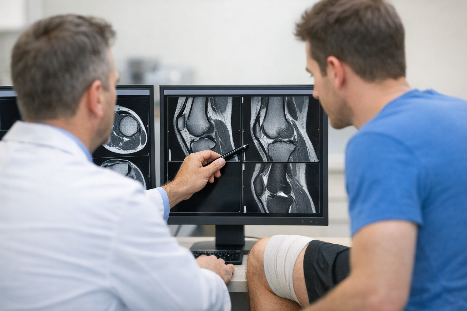
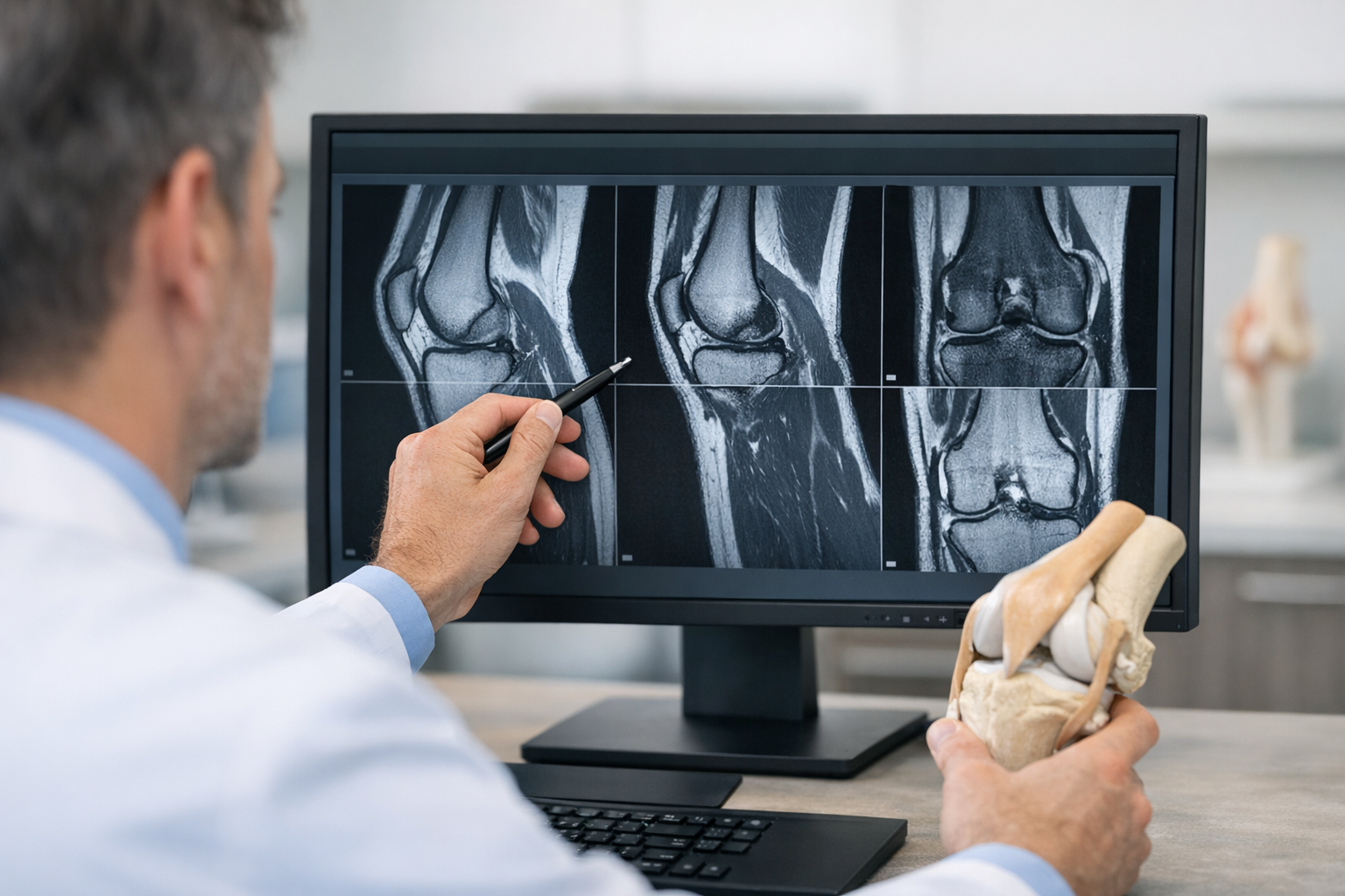
4. Imaging pitfalls: MRI false negatives and the “hidden accomplices”
MRI is valuable for confirming ACL injury and, critically, for assessing meniscus, cartilage, bone bruising, and multi-ligament patterns that shape management. However, clinicians still encounter the MRI false negative ACL tear problem, particularly with partial tears, suboptimal imaging conditions, or when the key issue is functional instability rather than a clearly visualized discontinuity.
Two practical implications:
- If MRI is reported as intact but the patient has recurrent giving way or exam-supported instability, treat function as data and re-triangulate history, re-exam, and imaging review rather than closing the case.
- Look beyond the ACL: associated injuries can drive symptoms and may coexist with subtle ACL deficiency.
For example, meniscal ramp lesions are frequently discussed in ACL reconstruction populations; their prevalence, imaging, and arthroscopic characterization have been reported in large cohorts such as Thaunat et al. (2021). The clinical point is not to assume a ramp lesion from symptoms alone, but to remember that meniscal pathology can contribute to persistent pain, mechanical symptoms, and perceived instability, complicating an ACL tear without pop narrative.
When the imaging story does not match the clinical story, a structured comparison of modalities can help align expectations: MRI vs arthrometer discussion is useful for explaining why imaging and functional assessment can disagree without implying that MRI is unnecessary.
5. When to escalate: objective laxity testing, ultrasound, and borderline pathways
Escalation is not “more tests for everyone.” It is targeted testing for patients whose function, sport demands, or exam uncertainty makes a false reassurance costly.
5.1 A practical escalation checklist
Consider escalation when any of the following are present:
- Recurrent giving way in sport, even with modest pain
- Inconsistent findings across visits, or pain-limited early exam that never gets repeated
- High-demand pivoting athlete with equivocal manual tests
- Discordance between MRI report and functional instability
In these scenarios, instrumented knee laxity testing may help quantify side-to-side instability and add functional information that complements clinical exam and MRI, particularly in equivocal cases, using tools such as the GNRB arthrometer or the Dyneelax arthrometer.
Keep the positioning clinically responsible: objective laxity data can support decision-making and follow-up planning, but MRI remains complementary and is typically needed to assess associated meniscal, chondral, and osseous injuries and for pre-operative planning when reconstruction is considered. If you want a broader rationale for standardizing measurement in ambiguous knees, see objective knee examination in orthopaedics.
5.2 Ultrasound and pediatric considerations
Dynamic ultrasound approaches are being explored as adjuncts for ACL assessment; a cadaveric diagnostic accuracy and reliability framework is described by Sakakibara et al. (2025). In practice, availability, operator expertise, and local validation determine whether this is a realistic pathway, but it can be part of “conditional escalation” where MRI access is delayed and clinical uncertainty is high.
Children and adolescents deserve extra caution: baseline laxity, communication limits, and different injury patterns can blur early findings. A focused review of pediatric cruciate ligament injury considerations is provided in Muenchgesang et al. (2026).
If the concern is “borderline MRI with ongoing instability,” align your follow-up sequence to a defined pathway: the workflow page provides a structured approach that keeps imaging and functional testing complementary.
For clinicians implementing measurement more broadly, keep the process consistent and auditable: quantitative laxity testing explains how objective side-to-side differences can be recorded and trended over time without overstating what any single number means.
6. Closing: key takeaways and next steps
The highest-risk scenario for delay is not the dramatic noncontact collapse. It is the athlete who finishes practice, presents later with vague pain, and is labeled “sprain” despite a pivot mechanism. That is where ACL tear without pop becomes a diagnostic trap.
- Do not over-weight the pop. Mechanism and effusion timing often carry more decision value.
- Re-exam is a strategy. If guarding blocks your assessment, schedule and document a timed re-check.
- Test rotation, not just translation. A meaningful pivot shift can be the key clue.
- Respect discordance. If function and exam suggest instability, reconsider even with reassuring imaging.
- Escalate conditionally. Use MRI for associated injuries and planning, and consider objective instability quantification when manual tests or MRI are equivocal.
If you are managing ongoing instability after an initially quiet injury, document the possibility of ACL tear without pop, protect the patient from pivoting during the diagnostic window, and close the loop with a defined follow-up plan to avoid another missed ACL rupture.
Clinical references (PubMed)
1) 2022 – Dietvorst et al. – Diagnostic values of history taking, physical examination and KT-1000 arthrometer for suspect anterior cruciate ligament injuries in children and adolescents: a prospective diagnostic study. – BMC Musculoskelet Disord – DOI: 10.1186/s12891-022-05659-1 – PMID: 35883084 – PubMed
2) 2025 – Sakakibara et al. – Diagnostic Accuracy and Reliability of Dynamic Handheld Ultrasound Testing in Detecting Anterior Cruciate Ligament Tears: A Cadaveric Study. – Arch Bone Jt Surg – DOI: 10.22038/ABJS.2025.87885.3980 – PMID: 41509056 – PubMed
3) 2021 – Thaunat et al. – Ramp Lesion Subtypes: Prevalence, Imaging, and Arthroscopic Findings in 2156 Anterior Cruciate Ligament Reconstructions. – Am J Sports Med – DOI: 10.1177/03635465211006103 – PMID: 33881943 – PubMed
4) 2023 – Yañez-Diaz et al. – Multiclass Support Vector Machine improves the Pivot-shift grading from Gerdy’s acceleration resultant prior to the acute Anterior Cruciate Ligament surgery. – Injury – DOI: 10.1016/j.injury.2023.03.020 – PMID: 37003872 – PubMed
5) 2026 – Muenchgesang et al. – [Ligamentous knee injuries in children: Focus on cruciate ligaments]. – Unfallchirurgie (Heidelb) – DOI: 10.1007/s00113-026-01703-0 – PMID: 41995736 – PubMed