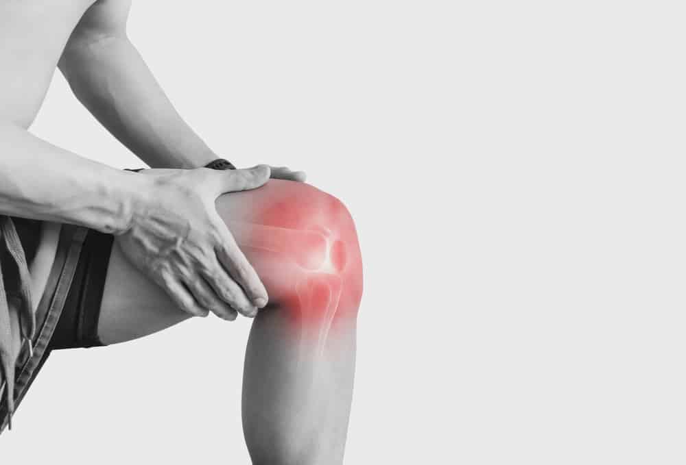
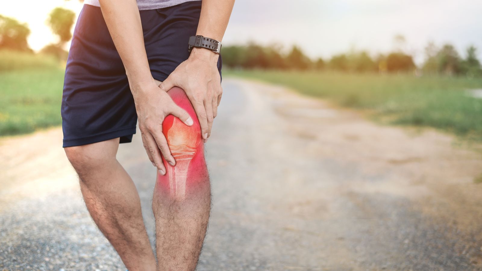
Introduction: Did I tear My ACL?
The Anterior Cruciate Ligament (ACL) plays a pivotal role in knee stability, and its injury can significantly impact mobility and quality of life. Diagnosing ACL injuries accurately is crucial for effective treatment and rehabilitation. In this article, we explore the various diagnostic methods for ACL injuries, with a special focus on the use of arthrometers, a groundbreaking tool that is revolutionizing the way we understand and treat these injuries.
1. Overview of ACL Injuries:
ACL injuries are common, particularly among athletes and active individuals. They can occur due to sudden stops, changes in direction, or direct impact. Symptoms often include pain, swelling, and a feeling of instability in the knee. Left undiagnosed or improperly managed, an ACL injury can lead to chronic knee problems and hinder a person’s ability to engage in physical activities.
2. Diagnostic Methods for ACL Injuries:
There are three primary methods for diagnosing an ACL injury: the use of an arthrometer/laximeter, physical examination, and MRI scans. Each method has its own set of advantages and limitations.
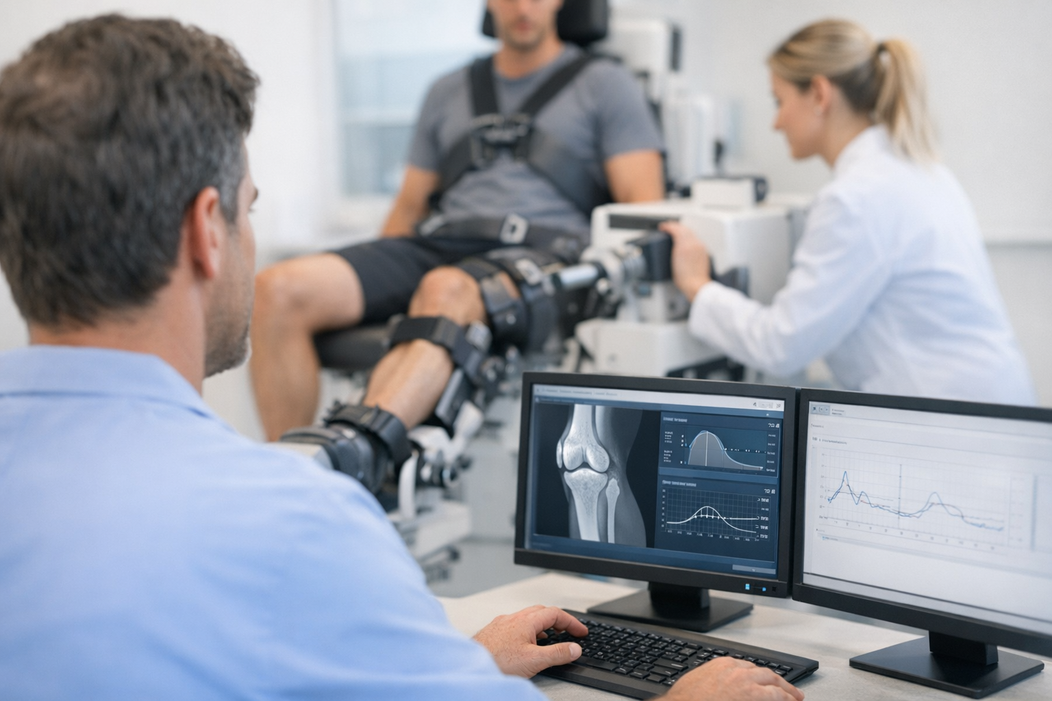
1) Arthrometer/Laximeter:
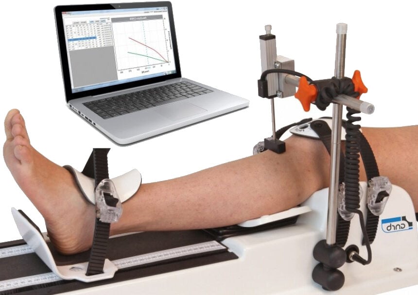
Arthrometers, like the KT-1000 and the more advanced GNRB (Comparison between KT1000 and GNRB: DOI: 10.1007/s00167-011-1869-2), offer precise and objective assessments of knee stability. These devices measure the displacement of the tibia under applied force, providing a clear picture of the ACL’s condition. Notably, arthrometers have been proven to be particularly effective in diagnosing partial ACL ruptures, often more efficiently compared to MRI. A significant study underscoring this point is titled “GNRB® laximeter with magnetic resonance imaging in clinical practice for complete and partial anterior cruciate ligament tears detection: A prospective diagnostic study with arthroscopic validation on 214 patients.”
Published in ‘The Knee’ journal, this study (DOI: 10.1016/j.knee.2023.03.017) provides robust clinical evidence of the arthrometer’s diagnostic superiority, especially in complex cases of partial ACL tears. The increasing prevalence of arthrometers in clinical settings can be attributed to their accuracy and reliability in diagnosing both complete and partial ACL injuries, as validated by such studies. In Korea, it was concluded the GNRB was superior to the Lachman test and Telos stress device (DOI: 10.5792/ksrr.17.014)
The Dyneelax® is currently the most advanced arthrometer developed by the company Genourob as it analyses the stability of the knee by applying both translation & rotation on the knee. A recent study, by Cojean et al. put forward its precision and reproducibility (DOI: 10.1016/j.medntd.2023.100254).
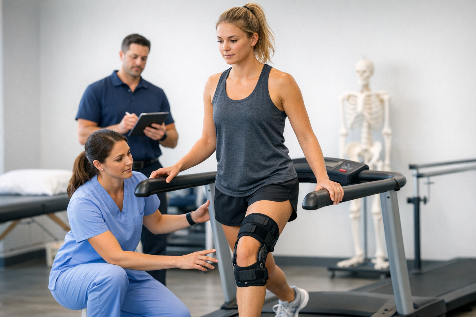
2) Physical Examination:
Physical examination remains a fundamental approach in the initial assessment of ACL injuries. It is a non-invasive method, usually performed by orthopaedic doctors or physiotherapists, to evaluate the integrity of the knee’s structures, including the ACL. This method comprises several specific tests, each designed to assess different aspects of knee function and stability.
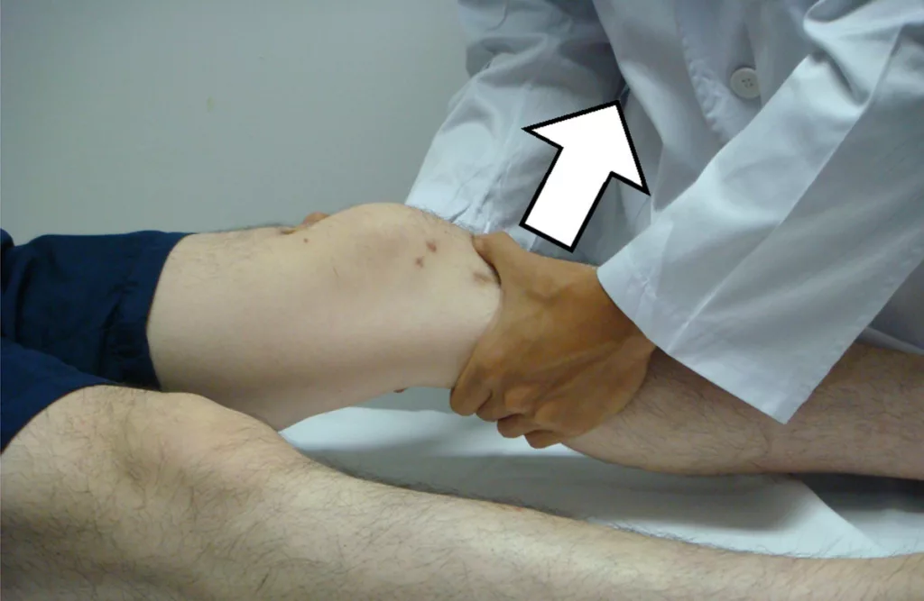
Lachman Test
This is one of the most sensitive tests for detecting ACL tears. The patient lies flat, and the knee is bent at a 20-30 degree angle. The examiner stabilizes the thigh while pulling the shin forward. Excessive forward movement of the shin indicates a potential tear in the ACL.
Anterior Drawer Test
In this test, the patient lies on their back with the knee flexed at 90 degrees. The examiner sits on the patient’s foot for stabilization and pulls the shin forward. Similar to the Lachman test, excessive forward movement suggests an ACL injury.
Pivot Shift Test
This dynamic test involves flexing and extending the knee while applying a valgus (inward) force and internal rotation to the lower leg. A positive test, indicated by a noticeable ‘shift’ or ‘clunk,’ suggests ACL insufficiency.
Jerk Test
The patient lies flat, and the knee is bent. The examiner then applies a lateral force to the knee while extending it. A jerk or snap during this motion can indicate an ACL injury.
While these clinical physical maneuvers are essential in the diagnostic process, their accuracy can vary based on subjective factors. These include the clinician’s experience, the patient’s muscle relaxation, and inherent knee variability. Each test’s sensitivity and specificity can differ, and often, a combination of tests is employed for a more accurate diagnosis.
In recent years, there has been an emphasis on refining physical examination techniques and training clinicians to improve diagnostic accuracy. Despite advancements, however, the intrinsic subjectivity and variability of physical examination mean that it is often used in conjunction with more objective diagnostic methods, such as arthrometry or MRI scans, especially in cases where the physical examination results are inconclusive or when a more detailed assessment of knee stability is required.
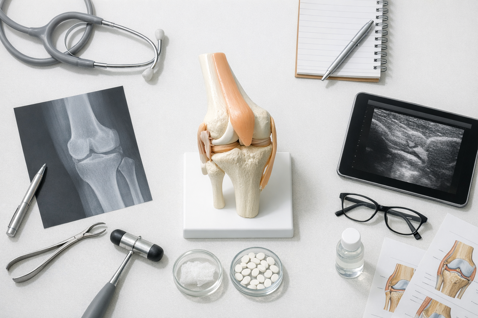
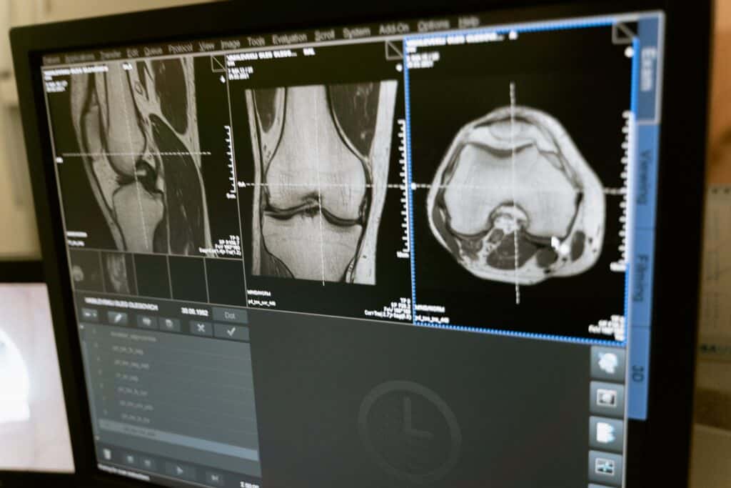
3) Magnetic Resonance Imaging:
Magnetic Resonance Imaging (MRI) plays a critical role in the diagnosis of ACL injuries, particularly in complex cases where a detailed view of soft tissues is required. MRI scans use strong magnetic fields and radio waves to create detailed images of the structures inside the knee, including bones, cartilage, and ligaments like the ACL.
Advantages of MRI in ACL Diagnosis:
- High-Resolution Images: MRI provides high-resolution images, offering a detailed view of the ACL and surrounding structures. This level of detail is crucial for assessing the extent of the injury.
- Non-Invasive: As a non-invasive imaging technique, MRI poses minimal risk to the patient and does not involve exposure to ionizing radiation.
- Comprehensive Evaluation: MRI can identify additional knee injuries that often accompany ACL tears, such as meniscal tears, bone bruises, or damage to other ligaments.
Limitations and Challenges:
- Interference by Blood: In acute injuries, the presence of blood or fluid in and around the ACL can obscure the view, making it challenging to determine the extent of the ligament’s damage.
- Static Nature of the Test: Since MRI scans are conducted with the patient lying still, they cannot assess the knee’s dynamic stability. This limitation means that the functional impact of the ACL injury might not be fully appreciated.
- Cost and Accessibility: MRI scans are relatively expensive and may not be readily available in all healthcare settings, limiting their use in certain populations or regions.
4) Detailed Focus on Arthrometer/Laximeter:
Arthrometers have significantly transformed the landscape of ACL diagnostics. Offering a precision and objectivity that surpass traditional methods, they have become essential tools in accurately determining ACL injuries. The GNRB arthrometer is particularly noteworthy; it applies varying forces to the knee, simulating the anterior drawer test. This approach generates detailed compliance curves of the ACL, providing vital insights into whether the ligament is partially or completely torn and the severity of such tears. This precise information is invaluable for planning effective treatment strategies.
Arthrometers are especially adept at identifying partial ACL ruptures, which are often more challenging to diagnose with other methods (GNRB Vs MRI: DOI: 10.1016/j.knee.2023.03.017). Their ability to measure subtle variations in knee stability makes them superior in detecting these less obvious injuries.
Complementary to MRI: While arthrometers offer a more precise and objective assessment, especially in the initial stages of diagnosis, they are not intended to replace MRI scans entirely. Instead, arthrometers serve as a less expensive, initial diagnostic tool for assessing the ACL. Once an ACL tear is confirmed through arthrometer testing, an MRI scan is still recommended for a comprehensive view of the knee. This approach ensures cost-effective testing, particularly beneficial for patients without a torn ACL, avoiding the higher costs of an MRI when unnecessary.
Synergy with Physical Examinations and MRI: Arthrometers, physical examinations, and MRI scans each play a distinct role in the diagnostic process. Arthrometers are particularly valuable when traditional methods yield inconclusive results or when a more detailed analysis of knee stability is necessary.
Guidance for Patients and Surgeons: Patients experiencing knee pain or instability are encouraged to discuss the option of using an arthrometer with their healthcare provider. For surgeons and clinicians, integrating arthrometers into the diagnostic process can significantly enhance the accuracy of ACL assessments, leading to improved patient outcomes and more informed treatment decisions.
Conclusion
The advent of arthrometers like the GNRB/Dyneelax has marked a significant advancement in the field of ACL diagnostics. By providing precise and objective assessments, especially in the challenging detection of partial ACL ruptures, these devices have filled a critical gap in the initial diagnostic process. While they do not aim to replace MRI scans, arthrometers offer a more cost-effective and efficient method for early assessment of ACL injuries. When used in conjunction with MRI scans and physical examinations, arthrometers contribute to a more comprehensive and accurate diagnostic approach, ultimately leading to better-informed treatment decisions and improved patient outcomes.
As we continue to embrace these technological advancements, the future of ACL diagnosis and treatment looks promising. By integrating tools like arthrometers into clinical practice, healthcare providers can ensure a higher standard of care for patients suffering from knee injuries, ensuring quicker, more accurate diagnoses and effective treatment plans tailored to individual needs.
Medical References (Link in DOI)
- Cojean, T. (2023). MRI vs. GNRB in ACL Injuries Detection: A Comparative Study. Journal of Orthopedic Research and Therapy (DOI: 10.1016/j.knee.2023.03.017).
- Cojean, T. (2023). Sensitivity, Repeatability, and Reproducibility Study with a Leg Prototype of a Recently Developed Knee Arthrometer Dyneelax. Journal of Biomechanics (DOI: 10.1016/j.medntd.2023.100254).
- Magdic, J. (2023). Intra-rater Reliability of the Knee Arthrometer GNRB. Journal of Clinical Orthopedics (DOI: 10.1016/j.jor.2023.03.016).
- Ryu, S.M. (2018). Diagnostic Tools for Acute Anterior Cruciate Ligament Injuries: A Comparative Analysis. Korean Journal of Sports Medicine (DOI: 10.5792/ksrr.17.014).
- Nouveau, S., Robert, H., & Viel, T. (2017). ACL Grafts Compliance During Time: Influence of Early Solicitations on the Final Stiffness of the Graft after Surgery. Journal of Orthopedic Research and Physiotherapy (DOI: 10.24966/ORP-2052/100035).
- Collette, M., Courville, J., Forton, M., & Gagnière, B. (2012). Objective Evaluation of Anterior Knee Laxity: Comparison of the KT-1000 and GNRB Arthrometers. Knee Surgery, Sports Traumatology, Arthroscopy (DOI: 10.1007/s00167-011-1869-2)