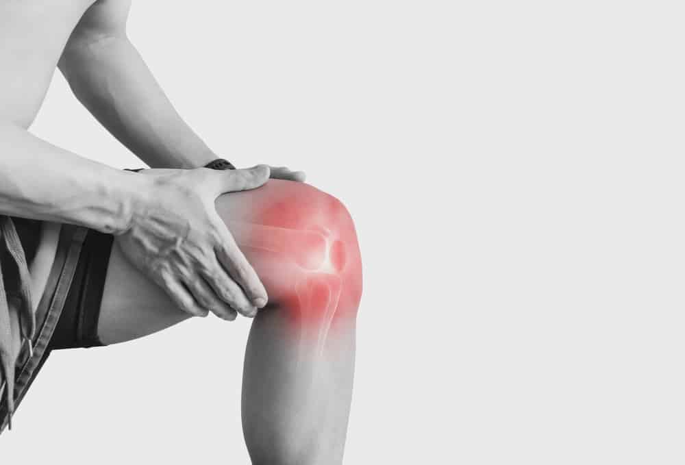
In ACL evaluation, the problem is often not whether laxity exists, but how reliably it is measured. That is where robotic knee laxity testing becomes clinically relevant. By standardizing load application during anterior tibial translation testing, clinicians can reduce one of the major weaknesses of manual exams: variable force. For orthopaedic surgeons, sports physicians, physiotherapists, and researchers, this matters because controlled loading may improve consistency, clarify borderline findings, and strengthen interpretation of instrumented knee laxity testing when the Lachman, pivot shift, and imaging do not fully align.
This guide explains why controlled force matters in ACL diagnosis, how to interpret objective dynamic laxity measurement, and where robotic approaches fit alongside MRI and standard examination rather than instead of them.
1. Why robotic knee laxity testing changes the diagnostic conversation
robotic knee laxity testing matters because ligament assessment is force-dependent. If the examiner applies different amounts of anterior force from one test to the next, the measured displacement can change even when the knee has not. That simple issue affects side-to-side comparison, longitudinal follow-up, and the ability to distinguish a partial injury from a functionally unstable ACL-deficient knee.
Traditional manual tests remain essential. The Lachman test, anterior drawer, and pivot shift are still central to ACL assessment, and they should not be sidelined. However, examiner feel is not the same as quantified loading. A more objective framework can help translate subjective impressions into reproducible values.
In practical terms, controlled force ACL diagnosis aims to answer three linked questions:
- How much displacement occurs?
- At what applied force does it occur?
- How does the involved knee compare with the contralateral side?
That is why robotic knee laxity testing is not just about a number. It is about obtaining a standardized force-displacement response that may better reflect functional instability than an uncontrolled push-pull exam alone.
Clinicians who already rely on the Lachman and pivot shift can use quantified data as an extension of bedside reasoning, especially when deciding whether a subtle finding is clinically meaningful, as discussed in this article on standard exams.
2. Controlled force and the mechanics of anterior tibial translation testing
ACL insufficiency is commonly assessed through anterior tibial translation testing. Yet anterior translation is not a fixed trait. It depends on positioning, hamstring guarding, tibial rotation, device fixation, and critically, the magnitude and rate of the applied load.
With uncontrolled testing, two experienced examiners may both identify laxity but generate different displacement values because they deliver different force profiles. In contrast, robotic knee laxity testing and automated arthrometer ACL platforms are designed to apply and record force in a standardized way, reducing one source of measurement noise.
2.1 Why force standardization matters
Standardized loading may improve interpretation in at least four situations:
- Acute ACL injury, when pain and guarding limit confidence in manual testing
- Partial or borderline ACL tears, where gross instability may be absent
- Postoperative follow-up, where small differences matter over time
- Research settings, where reproducibility is essential
The value is not only in documenting an ACL laxity side-to-side difference, but in understanding the load-response behavior that produces that difference. This is one reason force-response analysis can be more clinically informative than displacement alone, particularly when considering stiffness and compliance patterns.
Importantly, robotic knee laxity testing complements MRI rather than replacing it. MRI remains necessary in many cases to assess meniscal tears, cartilage injury, bone bruising, and preoperative anatomy. What quantified testing adds is functional and dynamic information that static imaging may not fully capture.
2.2 A short decision aid for clinicians
- Start with history, mechanism, swelling, and standard examination.
- If findings are obvious, proceed with clinician-led management and imaging as indicated.
- If the exam is limited, equivocal, or inconsistent with MRI, consider robotic knee laxity testing or other objective measures.
- Interpret results using force level, displacement pattern, and side-to-side comparison, not a single isolated value.
- Use MRI as a complementary tool, especially when associated injury or surgical planning is relevant.
This approach is especially useful in borderline MRI cases, where clinical uncertainty is often about function rather than structure alone.
3. Interpreting robotic knee laxity testing in real ACL diagnosis
The main clinical output of robotic knee laxity testing is not simply whether translation is increased, but whether the measured instability is consistent, side-specific, and mechanically plausible. Good interpretation requires context.
3.1 What to look for
In practice, interpretation often centers on:
- ACL laxity side-to-side difference
- The shape of the force-displacement curve
- End-point behavior and compliance
- Reproducibility across repeated trials
- Concordance with Lachman, pivot shift, symptoms, and MRI
That is why robotic knee laxity testing is best understood as part of a broader quantified instability assessment rather than a standalone verdict. Side-to-side analysis is often more clinically useful than an isolated raw value, particularly in patients with generalized laxity or variable baseline looseness. For a focused discussion of interpretation, see this guide on comparison.
When imaging and function diverge, objective testing may help narrow uncertainty. A patient with a structurally abnormal ACL on MRI but minimal functional asymmetry may differ clinically from a patient with subtle imaging findings and marked dynamic instability. This is also why MRI and arthrometric testing are often most informative when used together, as outlined in MRI comparison.
Relevant postoperative literature also reinforces the role of objective laxity measurement as an outcome tool. For example, Abram et al. (2026) reported no differences in objective knee laxity measurements between fixation strategies at 1 year after ACL reconstruction, illustrating how standardized laxity metrics can support comparative surgical evaluation. Similarly, Andre et al. (2026) examined outcomes after combined ACL and ALL reconstruction techniques, showing how objective postoperative assessment remains relevant when comparing reconstruction constructs.
3.2 Common pitfalls
Even with robotic knee laxity testing, interpretation is not automatic.
- Guarding can still affect readings
- Poor positioning can alter translation values
- Hyperlax patients need thoughtful contralateral comparison
- Rotational instability may not be fully captured by sagittal translation alone
Clinicians should also remember that some patients complain primarily of giving way during cutting or pivoting rather than straight-line translation symptoms. In those cases, sagittal instrumented knee laxity testing is helpful, but it may not tell the whole story.
4. Rotational instability, pivot shift, and what controlled force cannot fully answer
ACL deficiency is not purely an anterior translation problem. It is also a rotational instability problem. That distinction matters, because a patient may have measurable sagittal laxity and relatively limited pivot symptoms, or the reverse.
robotic knee laxity testing can strengthen ACL diagnosis by improving consistency in anterior loading, but it does not eliminate the need for rotational assessment. The pivot shift still carries major clinical relevance because it reflects dynamic subluxation behavior more directly than simple linear displacement.
So why does controlled force still matter in a rotationally complex condition? Because a reliable anterior translation baseline improves the quality of the whole assessment. When sagittal laxity is documented objectively, clinicians can better separate:
- clear ACL insufficiency with concordant findings
- subtle laxity with prominent rotational symptoms
- borderline translation with possible secondary stabilizer involvement
- postoperative residual looseness versus subjective apprehension
In this sense, robotic knee laxity testing helps structure the decision process, even if it does not replace the pivot shift. It can be particularly useful when considering whether apparent instability may reflect combined pathology involving the anterolateral complex, meniscus, or posterolateral structures.
Objective metrics also matter in early follow-up research. Forelli et al. (2025) evaluated early postoperative anterior knee laxity together with arthrogenic muscle inhibition and kinesiophobia, highlighting how laxity data should be interpreted alongside neuromuscular and patient-reported factors rather than in isolation.
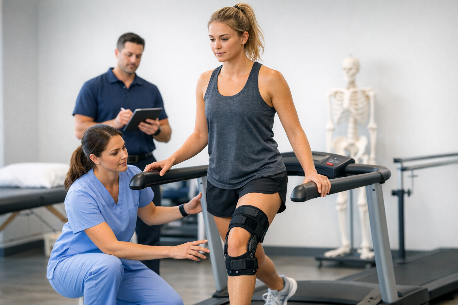
5. Where device-based testing fits in contemporary practice
When discussing robotic knee laxity testing, the core clinical question is not brand preference. It is whether the system can deliver objective dynamic laxity measurement with controlled force, repeatability, and meaningful side-to-side interpretation.
Examples of current device-based approaches include the GNRB arthrometer and the Dyneelax arthrometer, both of which are used in the context of quantified ACL assessment rather than as substitutes for MRI or clinician examination.
A clinically relevant GNRB arthrometer study highlighted greater measured knee laxity in ACL-deficient patients with lateral meniscal tears, reinforcing the value of standardized anterior translation assessment in complex injury patterns. A recent Dyneelax arthrometer study also reflects growing interest in technology-enabled quantification during ligament reconstruction evaluation.
The quality of any automated arthrometer ACL workflow still depends on protocol discipline. Reproducibility improves when patient setup, calibration, force progression, and operator training are standardized. That is one reason the issue of the learning curve remains relevant, even for highly standardized systems.
Device-specific reliability also remains an active research topic. Saito et al. (2026) assessed anterior tibial translation and device-specific reliability of the KNEELAX3 arthrometer in healthy women, reminding clinicians that reliability is device-dependent and population-dependent. Broader biologic factors can also affect ligamentous laxity over time, as seen in Bulguroglu et al. (2026), although that study addressed postpartum ligamentous recovery rather than ACL diagnosis directly.
For clinicians comparing available platforms, this overview of device comparison may help frame practical differences without losing sight of the larger diagnostic purpose.
6. Key takeaways and next steps
robotic knee laxity testing is most valuable when the force applied to the knee needs to be measured, standardized, and reproduced. In ACL diagnosis, controlled loading can improve confidence in anterior tibial translation testing, support interpretation of ACL laxity side-to-side difference, and refine understanding of objective dynamic laxity measurement.
Its main strengths are consistency and quantification. Its main limitation is that it cannot capture the entire complexity of ACL-related rotational instability on its own.
Practical next steps for clinicians:
- Use robotic knee laxity testing as a complement to history, manual examination, and MRI
- Prioritize side-to-side interpretation over isolated displacement values
- Consider force-response behavior, not just end translation
- Be cautious in partial tears, generalized laxity, and combined injuries
- Integrate findings with pivot shift, symptoms, and associated structural pathology
In suspected partial ACL tears or equivocal MRI cases, robotic knee laxity testing may offer added diagnostic value by quantifying functional instability that imaging does not fully show. MRI, however, remains complementary and is typically needed to assess meniscal, cartilage, and bony injury, as well as for preoperative planning when reconstruction is considered.
Ultimately, robotic knee laxity testing is not about replacing clinical judgement. It is about making ligament assessment more controlled, more transparent, and potentially more reproducible in the cases where precision matters most.
Clinical references (PubMed)
1) 2026 – Abram et al. – No differences in objective knee laxity measurements or patient-reported outcome measures between fixed- and adjustable-loop suspensory fixation in anterior cruciate ligament reconstruction: 1-year results from the GAP study, a prospective, double-blinded, randomized trial. – J ISAKOS – DOI: 10.1016/j.jisako.2026.101123 – PMID: 42044694 – PubMed
2) 2026 – Andre et al. – Comparable outcomes following combined ACL and ALL reconstruction using a 1-strand versus 2-strand back-and-forth technique: Propensity score matched study. – J Exp Orthop – DOI: 10.1002/jeo2.70658 – PMID: 41799400 – PubMed
3) 2025 – Forelli et al. – Early Postoperative Evaluation of Arthrogenic Muscle Inhibition, Anterior Knee Laxity, and Kinesiophobia After ACL Reconstruction: A Cross-Sectional Observational Study. – Healthcare (Basel) – DOI: 10.3390/healthcare13131481 – PMID: 40648507 – PubMed
4) 2026 – Bulguroglu et al. – Effect of continued prenatal Pilates until childbirth on postpartum ligamentous recovery: a longitudinal follow-up of a randomized controlled trial. – BMC Pregnancy Childbirth – DOI: 10.1186/s12884-026-09183-1 – PMID: 42067815 – PubMed
5) 2026 – Saito et al. – Anterior tibial translation and device-specific reliability of the KNEELAX3 arthrometer in healthy women. – J Orthop Sci – DOI: 10.1016/j.jos.2026.05.010 – PMID: 42225498 – PubMed