A combined ACL MCL injury is not simply an ACL tear plus a painful medial sprain. In many athletes and active patients, the medial side determines whether the knee behaves like an isolated sagittal-plane problem or a more complex instability pattern with medial knee instability, valgus overload, and persistent rotation. That is why careful assessment of medial side knee stability matters early. If the medial structures are under-recognized, clinicians may underestimate residual laxity, misread the pivot shift, or choose treatment pathways that do not fully address function. For orthopaedic surgeons, sports physicians, physiotherapists, and researchers, the key issue is not only diagnosis, but how the medial side changes decision-making.
1. Why combined ACL MCL injury is more than a “double ligament” problem
A combined ACL MCL injury changes knee mechanics in ways that are easy to simplify and hard to manage if the medial side is not mapped properly. The superficial MCL, deep MCL, posterior oblique ligament, capsule, and posteromedial structures all contribute to restraint patterns that interact with ACL deficiency.
Clinically, this means an ACL and MCL tear may present with more than anterior translation. Depending on the injured structures, the patient may show valgus laxity ACL injury patterns, excess external rotation, or signs consistent with anteromedial rotatory instability. In practice, that can influence the pivot shift, perceived giving way, graft stress after ACL reconstruction, and return-to-sport confidence.
The medial side is particularly important because the medial structures help control both translational and rotational behavior. This is one reason the MCL role in ACL stability has moved from a secondary consideration to a core element of modern instability assessment.
Recent literature supports the need for nuanced management rather than one-size-fits-all algorithms. In combined injuries requiring operative consideration, Chung et al. (2026) evaluated one-stage versus two-stage ACL reconstruction with concomitant MCL surgery, highlighting that timing and strategy remain active clinical questions rather than settled doctrine.
2. How the medial side alters instability patterns in combined ACL MCL injury
In a combined ACL MCL injury, the medial side can amplify instability in three main domains:
- Valgus control in near extension and at 20 to 30 degrees
- Anterior restraint sharing, especially under dynamic load
- Rotational control, particularly anteromedial and pivot-related phenomena
When clinicians focus only on Lachman findings, they may miss the broader pattern of a combined knee ligament injury. A disrupted medial envelope can increase anterior tibial translation under functional conditions and may also worsen apparent rotational instability. This is why the pivot shift in some ACL-deficient knees reflects more than the lateral side alone.
Equally, not every medial lesion behaves the same way. A low-grade MCL injury with good healing potential may not have the same functional consequences as a high-grade tear with posteromedial involvement. If the posterior oblique ligament or capsule is involved, the pattern can look less like a routine ACL and MCL tear and more like a rotationally relevant medial failure complex.
2.1. Why anteromedial rotation matters
Anteromedial rotatory instability is a useful concept because it explains why some knees feel unstable despite seemingly acceptable isolated ACL metrics. If the medial side does not restrain rotation and translation effectively, the patient may report cutting instability, valgus collapse sensations, or recurrent episodes during deceleration.
The practical implication is simple: in combined ACL MCL injury, the examiner should ask not only “how loose is the ACL?” but also “which medial restraints are failing, and in which plane?” This is well aligned with more comprehensive objective exam frameworks.
3. Clinical assessment: what to test, when to worry, and common pitfalls
The first challenge in combined ACL MCL injury is separating pain inhibition from true instability. Acutely, guarding can suppress findings. Later, a healing MCL can mask residual laxity if the examination is too limited or performed only in one position.
3.1. Core examination priorities
A focused assessment should usually include:
- Lachman and anterior drawer
- Valgus stress testing in extension and at 20 to 30 degrees
- Pivot shift, if tolerated and safe
- Assessment for posteromedial tenderness or opening
- Comparison with the contralateral knee
Structured valgus stress testing helps define grade, location, and whether the apparent MCL lesion behaves like an isolated injury or part of a larger instability pattern. In a combined ACL MCL injury, the amount of medial opening is only one part of interpretation. The key is whether the opening is accompanied by rotational symptoms, persistent effusion, apprehension, or a high-grade pivot phenomenon.
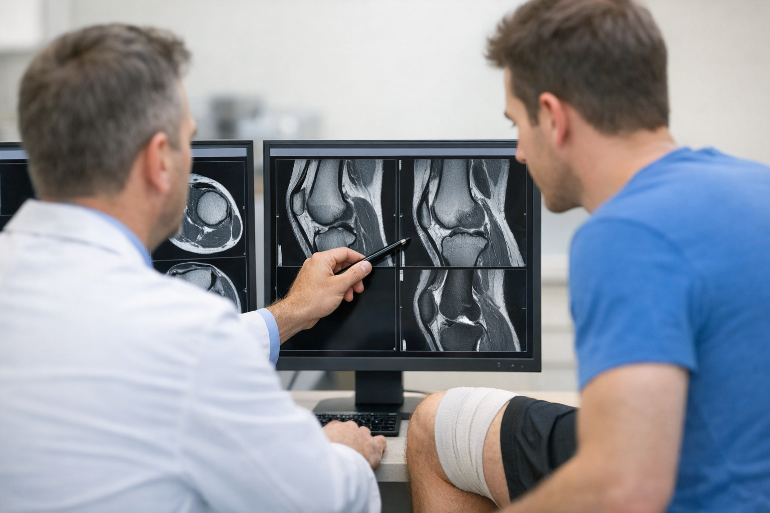
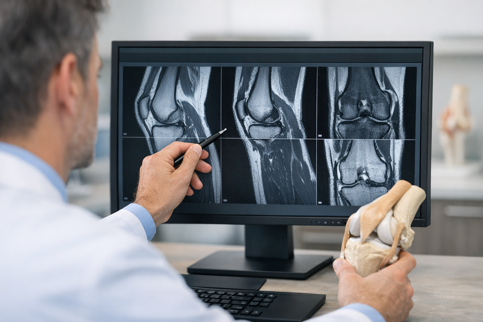
Clinicians should also remember that MRI remains important. Imaging can define meniscal, chondral, bony, and capsuloligamentous injury, and it is typically needed for surgical planning when reconstruction is considered. In equivocal cases, especially when MRI findings are borderline for partial ACL injury, functional instability may still need clarification through clinical and objective testing rather than imaging alone. The workflow described for borderline MRI cases reflects this complementary approach.
For background concepts that often shape interpretation, knee laxity is best understood as a measured mechanical finding that may or may not match patient-reported instability.
3.2. Common pitfalls
- Assuming every MCL injury will heal functionally enough to protect an ACL graft
- Ignoring extension valgus opening, which may suggest more extensive medial involvement
- Underestimating the significance of a high-grade pivot in a medial-sided injury
- Relying on MRI wording alone without correlating examination findings
These pitfalls are especially relevant in combined ACL MCL injury, where the line between nonoperative medial care and surgical medial treatment can be narrow.
4. Decision-making in combined ACL MCL injury: nonoperative, one-stage, or staged?
Management of combined ACL MCL injury is rarely binary. The main decision variables are injury grade, tissue quality, chronicity, associated lesions, alignment, activity goals, and whether instability persists after early MCL treatment.
A short decision aid can help:
- Confirm the pattern – Is this isolated ACL plus low-grade MCL, or a more complex medial-sided injury?
- Grade the medial lesion – Is there clinically meaningful valgus or rotational instability?
- Reassess after acute settling – Has medial function improved enough to support isolated ACL reconstruction, if needed?
- Escalate if instability persists – Persistent valgus, rotational laxity, or clear posteromedial insufficiency may justify combined treatment.
In operative pathways, the question is often whether to perform ACL reconstruction alone, ACL reconstruction with concurrent medial surgery, or a staged strategy. Chung et al. (2026) specifically addressed one-stage versus two-stage ACL reconstruction with concomitant MCL surgery in this setting, reinforcing that treatment timing should be individualized.
For the medial side itself, Lack et al. (2026) reported a systematic review suggesting comparable outcome scores for MCL reconstruction and repair in grade III injuries, with lower complication rates after repair at 2-year follow-up. That does not mean repair is always preferable, but it supports tissue- and context-based decision-making.
When the injury is not a simple ACL plus MCL pattern but part of a broader combined knee ligament injury, the considerations become more complex. The review by Hantes et al. (2026) is useful in framing what general orthopaedic surgeons should recognize in multiligament presentations.
For readers comparing medial reconstructive strategies in relation to anterior and rotational control, this discussion of medial-side reconstruction provides added context.
5. Where objective laxity testing may help in combined ACL MCL injury
Because a combined ACL MCL injury often behaves in more than one plane, selective use of objective or dynamic laxity testing may help quantify side-to-side instability and complement the clinical exam and MRI, especially when the examination is limited by guarding or when residual functional instability is uncertain.
In practice, multi-axis approaches can be more informative than a single manual endpoint because they may better reflect how medial side knee stability and ACL restraint interact under load. A structured multi-axis workflow may be particularly relevant in suspected rotational or multiligament patterns.
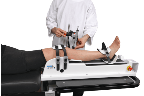
If instrumented assessment is being considered, GNRB testing and Dyneelax assessment may provide complementary quantitative information, but they do not replace MRI and should be interpreted within a clinician-led diagnostic pathway.
This matters in combined ACL MCL injury because some knees look “acceptable” on static imaging yet remain functionally unstable. Objective measurements can add context to whether persistent symptoms reflect anterior laxity, valgus contribution, or unresolved rotational control deficits.
6. Rehabilitation, return to sport, and why the medial side still matters later
Even after the acute treatment decision is made, combined ACL MCL injury should not be followed like a standard isolated ACL pathway. The medial side still matters during strength progression, change-of-direction work, and return-to-sport testing.
For professional athletes, return outcomes after combined injury may differ from isolated ACL patterns. Abdul et al. (2026) compared isolated ACL and combined ACL/MCL injuries in professional soccer and rugby players, focusing on return to play and career longevity. That type of evidence is clinically relevant because it reminds teams that timeline expectations may need adjustment when the medial side is involved.
In chronic follow-up or multiligament reconstructions involving the posteromedial side, longer-term data also matter. Chamundaiah et al. (2026) reported 10-year clinical and functional outcomes after acute MCL with postero-medial complex repair in multiligament knee injuries using a double shoelace repair technique, highlighting the importance of durable medial control in selected cases.
From a rehabilitation standpoint, progression should be based on function plus stability, not time alone. These resources on rehab milestones and return to sport are relevant when residual valgus or rotational concerns may change loading tolerance and readiness decisions.
In other words, a combined ACL MCL injury that looks calm in the clinic can still fail under sport-specific demand if medial restraint has not normalized sufficiently.
7. Key takeaways and next steps
The central message is that combined ACL MCL injury should be approached as a stability pattern, not just a list of torn structures. The medial side influences valgus control, anterior translation sharing, rotational behavior, and potentially ACL graft load. That is why careful grading, repeat examination, and context-specific treatment planning matter.
Key points:
- A combined ACL MCL injury may behave very differently from an isolated ACL tear.
- The MCL role in ACL stability becomes especially important when valgus opening, posteromedial pain, or rotational symptoms are present.
- Anteromedial rotatory instability and the pivot shift medial structures relationship should be actively considered.
- MRI is complementary and remains important for associated injuries and preoperative planning.
- Objective laxity testing may help quantify instability in selected cases, but it should support, not replace, clinician-led assessment.
Next steps are straightforward: define the exact medial injury pattern, correlate imaging with examination, reassess after the acute phase when needed, and decide whether the patient has an isolated ACL problem with a healing MCL or a true combined ACL MCL injury requiring broader stabilization strategy.
Clinical references (PubMed)
1) 2026 – Chung et al. – One-Stage Versus Two-Stage ACL Reconstruction with Concomitant MCL Surgery in Combined ACL and MCL Injuries: A Minimum 2-Year Follow-Up Study. – J Clin Med – DOI: 10.3390/jcm15020583 – PMID: 41598520 – PubMed
2) 2026 – Lack et al. – Comparable Outcome Scores for Medial Collateral Ligament Reconstruction and Repair in Isolated and Combined Grade III Injuries, with Lower Rates of Complication Following Repair at 2-year Follow-up: A Systematic Review. – J Knee Surg – DOI: 10.1055/a-2778-8771 – PMID: 41592587 – PubMed
3) 2026 – Abdul et al. – Comparison of Isolated ACL and Combined ACL/MCL Injuries in Professional Soccer and Rugby Players: Return to Play and Career Longevity Outcomes. – Am J Sports Med – DOI: 10.1177/03635465261448236 – PMID: 42212766 – PubMed
4) 2026 – Chamundaiah et al. – Acute Medial Collateral Ligament With Postero-Medial Complex Repair in Multiligament Knee Injuries Using a Novel ‘Double Shoelace Repair’ Technique: 10-Year Clinical and Functional Outcomes. – Cureus – DOI: 10.7759/cureus.102328 – PMID: 41755975 – PubMed
5) 2026 – Hantes et al. – Multiligament-injured knee: what the general orthopedic surgeon should know. – EFORT Open Rev – DOI: 10.1530/EOR-2026-0053 – PMID: 42065223 – PubMed