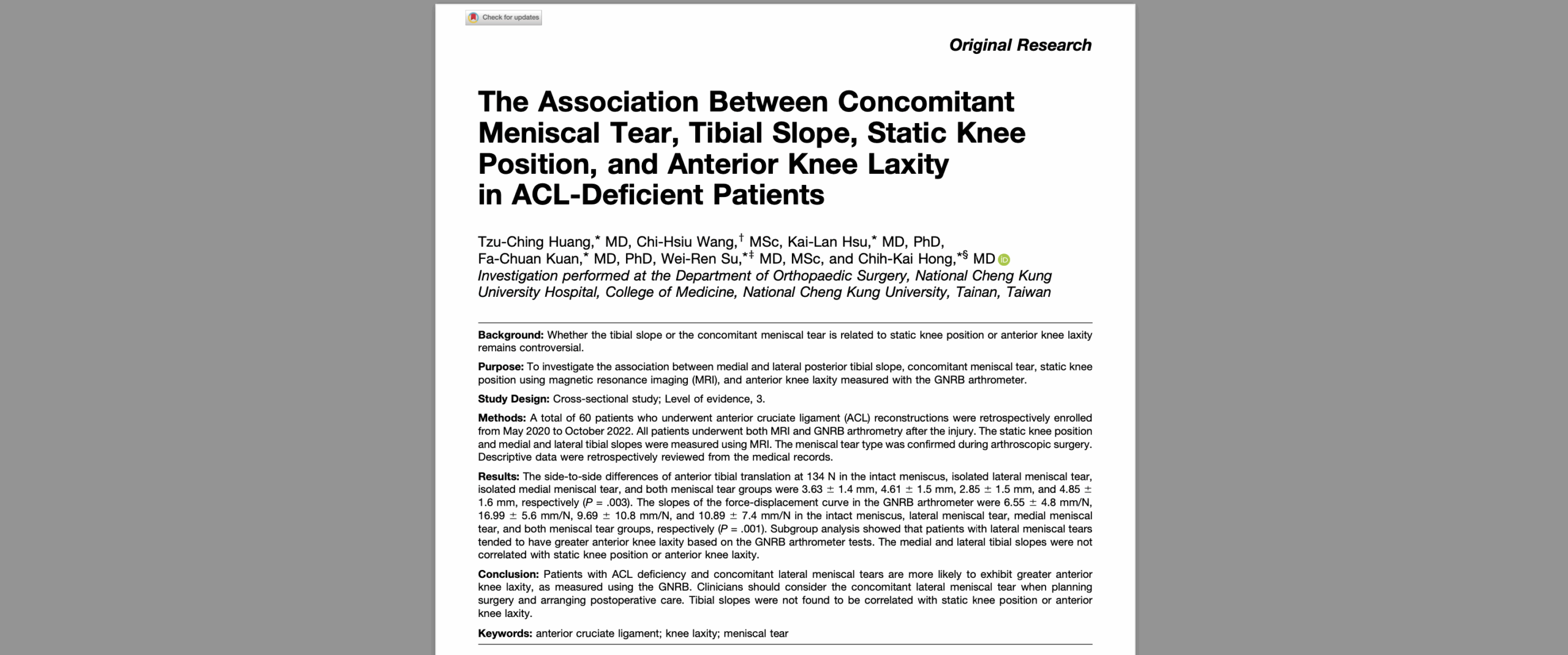
Knee laxity is a critical concern in the field of orthopedics, often arising from injuries to the ligaments, such as the anterior cruciate ligament (ACL). Accurate diagnosis and effective management of knee laxity are essential for preventing long-term damage and ensuring optimal patient outcomes. Knee laxity testing is a fundamental diagnostic process used to evaluate the stability and integrity of the knee joint.
In recent years, advances in medical technology have significantly improved the precision and reliability of these tests. Traditional methods, while useful, often lack the consistency and accuracy needed for definitive diagnosis. This is where robotic arthrometers come into play, offering a new standard in knee laxity testing.
Among the leading innovations are the GNRB and Dyneelax robotic arthrometers. These devices provide objective measurements and quantitative data that enhance the diagnostic process, making it possible to detect even subtle degrees of knee laxity. By integrating these advanced tools into clinical practice, orthopedic professionals can achieve more accurate assessments, leading to better treatment plans and improved patient outcomes.
In this article, we will explore the significance of the knee laxity test, delve into the traditional methods, and highlight the advantages of using GNRB and Dyneelax robotic arthrometers.
I. Understanding Knee Laxity

Knee laxity refers to the looseness or instability of the knee joint, which can lead to impaired function and discomfort. This condition typically arises when the ligaments that support the knee become stretched, torn, or otherwise damaged. The primary ligaments involved in maintaining knee stability include the anterior cruciate ligament (ACL), posterior cruciate ligament (PCL), medial collateral ligament (MCL), and lateral collateral ligament (LCL). When these ligaments are compromised, the knee can become unstable, leading to a range of symptoms including pain, swelling, and difficulty in movement.
Common causes of knee laxity include traumatic injuries, such as those sustained during sports activities or accidents. For example, a sudden change in direction, a direct blow to the knee, or a landing awkwardly from a jump can cause ligament tears. Other causes include repetitive strain from overuse, which can weaken the ligaments over time, and degenerative conditions like osteoarthritis, which can erode the structures supporting the knee.
There are different types of knee laxity, categorized based on the specific ligament affected. ACL injuries are among the most common, particularly in athletes involved in sports that require sudden stops and changes in direction. PCL injuries are often a result of a direct impact to the front of the knee. MCL and LCL injuries typically occur due to lateral forces applied to the knee.
Early diagnosis and treatment of knee laxity are crucial to prevent further damage and to restore knee function. Delayed treatment can lead to chronic instability, which increases the risk of additional injuries such as meniscal tears and cartilage damage. Accurate diagnosis typically involves a combination of physical examinations, imaging studies, and specialized tests such knee laxity testing. Timely intervention, whether through conservative management like physical therapy or surgical reconstruction, can significantly improve outcomes and help individuals return to their normal activities with reduced risk of long-term complications.
Understanding knee laxity and recognizing its symptoms promptly can make a substantial difference in the effectiveness of the treatment and the overall prognosis for the patient.
II. Traditional Methods of Knee Laxity Testing
Traditional knee laxity tests have been the cornerstone of diagnosing ligamentous injuries in the knee for many years. These manual tests are designed to evaluate the stability and integrity of the knee ligaments, particularly focusing on detecting any abnormal looseness or instability.
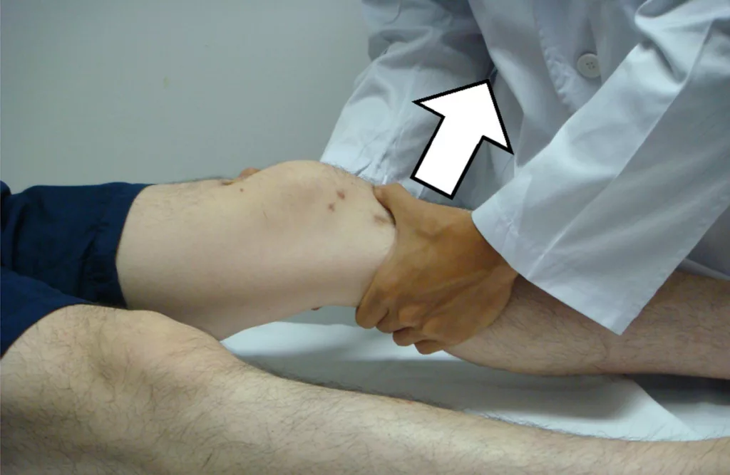
One of the most commonly used tests is the Lachman test, which is highly regarded for its sensitivity in diagnosing anterior cruciate ligament (ACL) injuries. During this test, the patient lies supine with the knee flexed at approximately 20-30 degrees. The examiner stabilizes the thigh while pulling the tibia forward to assess the degree of anterior translation. An increased forward movement of the tibia compared to the unaffected knee suggests an ACL tear.
Another widely used test is the Pivot Shift test, which also assesses ACL integrity. The patient is positioned supine, and the examiner applies a valgus force while internally rotating the tibia and flexing the knee. A positive test is indicated by a palpable clunk or shift as the tibia reduces on the femur, signifying anterolateral rotatory instability due to an ACL tear.
The Anterior Drawer test is another traditional method, where the knee is flexed to 90 degrees, and the examiner pulls the tibia forward while stabilizing the foot. This test, however, is less sensitive than the Lachman test and is often used in conjunction with other assessments.
Despite their widespread use, traditional knee laxity tests have several limitations and challenges. One major limitation is the reliance on the examiner’s experience and skill, which can lead to variability in the results. Factors such as patient muscle guarding, pain, and body habitus can also affect the accuracy of these tests. Moreover, these manual tests provide qualitative rather than quantitative data, making it difficult to precisely measure the degree of knee laxity.
Another challenge is that these tests may not be as reliable in the acute setting when swelling and pain can obscure the physical findings. Additionally, they can sometimes fail to detect partial ligament tears or subtle degrees of instability, leading to potential underdiagnosis.
In summary, while traditional knee laxity tests like the Lachman test and Pivot Shift test are essential tools in orthopedic practice, they are not without their limitations. The development of more advanced diagnostic tools, such as robotic arthrometers, aims to address these challenges by providing more consistent and objective measurements of knee laxity.
III. Advances in Knee Laxity Testing: Robotic Arthrometers
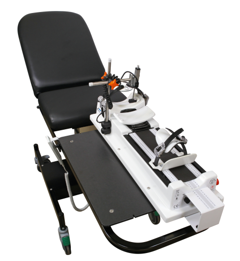
Advances in knee laxity testing have been significantly enhanced by the introduction of robotic arthrometers, which provide a new level of precision and reliability in diagnosing ligament injuries. Unlike traditional manual tests, robotic arthrometers utilize sophisticated technology to deliver objective, quantitative data regarding knee stability.
Robotic arthrometers operate by applying controlled forces to the knee joint and measuring the resultant displacement with high accuracy. This process minimizes the variability associated with manual tests and reduces the influence of examiner skill and patient factors such as muscle guarding or pain. The devices can consistently replicate testing conditions, ensuring that each assessment is comparable to the last, which is critical for tracking patient progress over time.
Studies have demonstrated the benefits of robotic arthrometers in enhancing knee ligament treatment. For instance, Cojean et al. (2023) found that robotic arthrometers are superior to MRI in detecting partial ACL ruptures, offering better sensitivity and specificity . Additionally, Guegan et al. (2024) highlighted that the Dyneelax arthrometer not only measures ACL laxity with precision but also evaluates the surrounding structures, providing a comprehensive assessment of knee stability and helping to plan for surgery precisely, such as choosing lateral extra-articular tenodesis (LET) surgery . Furthermore, Forelli (2023) showed how robotic arthrometers are instrumental in post-operative rehabilitation, allowing for personalized return-to-sport programs based on precise laxity measurements .
These advanced tools not only enhance diagnostic accuracy but also improve patient outcomes by enabling early and precise detection of ligament injuries. The data provided by robotic arthrometers can be used to guide personalized treatment plans, whether through conservative management or surgical options.
IV. Dynamic Knee Laxity Testing with Robotic Arthrometers: GNRB and Dyneelax
4.1 Detailed Description of the Devices
The GNRB and Dyneelax robotic arthrometers represent the pinnacle of modern technology in knee laxity testing. The GNRB focuses on applying controlled anterior translation forces to the tibia, providing precise measurements of knee anterior laxity.
In contrast, the Dyneelax not only measures anterior tibial translation but also assesses rotational laxity, capturing both internal and external tibial rotations. This dual functionality of the Dyneelax offers a comprehensive assessment of knee stability, addressing the limitations of traditional methods and providing a more detailed picture of knee joint mechanics.

4.2 How GNRB and Dyneelax Perform the Knee Laxity Testing
The GNRB device operates by positioning the patient’s knee in slight flexion (around 20 degrees) and applying a controlled anterior force while recording the displacement of the tibia relative to the femur. The precise force-displacement data is then analyzed to assess knee laxity. Similarly, the Dyneelax performs anterior translation tests but extends its capabilities to measure internal and external rotations by applying rotational forces to the tibia. This additional rotational assessment allows for a thorough evaluation of the knee’s ligamentous structures, particularly useful in complex cases where multiple structures may be involved.
4.3 Additional Case Studies or Research Supporting the Effectiveness of GNRB and Dyneelax
Several other studies have also validated the effectiveness of these devices. Kayla Smith et al. (2022) confirmed the reliability of the GNRB in measuring ACL stiffness and laxity, emphasizing its clinical utility . Additionally, Magdic (2023) reported excellent intra-rater reliability of the GNRB for measuring knee anterior laxity . The Dyneelax also demonstrated high reliability in both translational and rotational measurements, as shown by Nascimento et al. (2024) and another study from Cojean et al. (2023), underscoring its effectiveness in clinical settings.
Other significant studies include Klouche et al. (2015), which demonstrated the diagnostic value of the GNRB in complete ACL tears, and Jenny et al. (2017), which validated the GNRB for measuring anterior tibial translation. Nouveau et al. (2017) highlighted the influence of early solicitations on graft stiffness after surgery, emphasizing the importance of precise laxity measurements for effective rehabilitation. Pouderoux et al. (2019) discussed the increase in joint laxity and graft compliance over time, further supporting the need for accurate and consistent monitoring tools like GNRB and Dyneelax .
Conclusion
Advanced robotic arthrometers, such as the GNRB and Dyneelax, represent significant advancements in the field of knee laxity testing. These devices provide objective, quantitative data that dramatically improves the accuracy and reliability of diagnosing ligament injuries compared to traditional manual methods. The ability to detect subtle degrees of laxity and measure both translational and rotational knee movements allows for more comprehensive evaluations, leading to better-informed clinical decisions and more effective treatment plans.
The evidence from multiple studies, including those by Cojean et al. (2023), Guegan et al. (2024), and Forelli (2023), underscores the superior diagnostic capabilities and clinical benefits of these robotic arthrometers. By adopting GNRB and Dyneelax technologies, orthopaedic professionals can enhance diagnostic accuracy, facilitate early intervention, and improve patient outcomes through personalised treatment and rehabilitation plans.
Orthopaedic professionals are encouraged to integrate these advanced tools into their practice to leverage their full potential. The precision and consistency offered by robotic arthrometers not only improve patient care but also set a new standard in the diagnosis and management of knee injuries. Embracing these innovations is a critical step towards advancing orthopaedic diagnostics and ensuring the highest quality of patient care.
Medical References (Link Available)
- Cojean T, Batailler C, Robert H, Cheze L. (2023). GNRB® laximeter with magnetic resonance imaging in clinical practice for complete and partial anterior cruciate ligament tears detection: A prospective diagnostic study with arthroscopic validation on 214 patients. Knee, 42, 373-381. DOI: 10.1016/j.knee.2023.03.017
- Guegan, B., Drouineau, M., Common, H., & Robert, H. (2024). All the menisco-ligamentary structures of the medial plane play a significant role in controlling anterior tibial translation and tibial rotation of the knee: Cadaveric study of 29 knees with the Dyneelax® laximeter. Journal of Experimental Orthopaedics, 11, e12038. DOI: 10.1002/jeo2.12038
- Forelli F, Le Coroller N, Gaspar M, et al. (2023). Ecological and Specific Evidence-Based Safe Return To Play After Anterior Cruciate Ligament Reconstruction In Soccer Players: A New International Paradigm. IJSPT. Published online April 2, 2023. DOI: 10.26603/001c.73031
- Smith K, Miller N, Laslovich S. (2022). The Reliability of the GNRB® Knee Arthrometer in Measuring ACL Stiffness and Laxity: Implications for Clinical Use and Clinical Trial Design. Int J Sports Phys Ther, 17(6), 1016-1025. DOI: 10.26603/001c.38252
- Magdić M, Gošnak Dahmane R, Vauhnik R. (2023). Intra-rater reliability of the knee arthrometer GNRB® for measuring knee anterior laxity in healthy active subjects. Journal of Orthopaedics. DOI: 10.1016/j.jor.2023.03.016
- Nascimento, N., Kotsifaki, R., Papakostas, E., Zikria, B. A., Alkhelaif, K., Hagert, E., Olory, B., D’Hooghe, P., & Whiteley, R. (2024). DYNEELAX Robotic Arthrometer Reliability and Feasibility on Healthy and Anterior Cruciate Ligament Injured/Reconstructed Persons. Translational Sports Medicine, 2024(1), 1-5. DOI: 10.1155/2024/3413466
- Cojean, T., Batailler, C., Robert, H., Cheze, L. (2023). Sensitivity repeatability and reproducibility study with a leg prototype of a recently developed knee arthrometer: The DYNEELAX®. Medicine in Novel Technology and Devices, 19, 100254. DOI: 10.1016/j.medntd.2023.100254
- Klouche S, Lefevre N, Cascua S, Herman S, Gerometta A, Bohu Y. (2015). Diagnostic value of the GNRB® in relation to pressure load for complete ACL tears: A prospective case-control study of 118 subjects. Orthopaedics & Traumatology: Surgery & Research, 101(3), 297–300. DOI: 10.1016/j.otsr.2015.01.008
- Jenny J-Y, Puliero B, Schockmel G, Harnoist S, Clavert P. (2017). Experimental validation of the GNRB® for measuring anterior tibial translation. Orthopaedics & Traumatology: Surgery & Research. Received: 7 October 2016, Accepted: 30 December 2016 DOI: 10.1016/j.otsr.2016.12.011
- Nouveau, S., Robert, H., Viel, T. (2017). ACL Grafts Compliance During Time: Influence of Early Solicitations on the Final Stiffness of the Graft after Surgery. Journal of Orthopedic Research and Physiotherapy, 3(1), 035. DOI: 10.24966/ORP-2052/100035
- Pouderoux, T., Muller, B., Robert, H. (2019). Joint laxity and graft compliance increase during the first year following ACL reconstruction with short hamstring tendon grafts.. DOI: 10.1007/s00167-019-05711-z

