“Borderline” ACL findings on MRI can leave the team uncertain: the report suggests a sprain or partial disruption, yet the patient describes giving way and loss of trust in pivoting. In this gap, borderline ACL MRI instability testing aims to align structure with function by prioritizing clinical correlation and reproducible instability assessment. This is especially relevant when a partial ACL tear MRI pattern is reported, but the exam is subtle, painful, or inconsistent between assessors. The goal is not to overrule imaging, but to add functional evidence that supports a confident pathway, whether that is rehabilitation, bracing, return-to-sport progression, or surgical consultation based on instability, demands, and associated injuries.
1) Defining the “borderline” MRI ACL and when borderline ACL MRI instability testing is triggered
Borderline MRI language commonly includes “ACL sprain,” “partial tear,” “attenuation,” “mucoid change,” “indeterminate fibers,” or “near-complete tear.” Radiology is describing morphology at one timepoint. Clinicians are deciding if the knee is functionally unstable during cutting, deceleration, and rotation.
borderline ACL MRI instability testing is most useful when at least one of the following is true:
- Symptoms suggest instability (pivoting episodes, “shifts,” or apprehension) but the MRI is equivocal.
- Acute pain, swelling, or guarding makes exam quality variable.
- The report emphasizes partial disruption but the patient has high rotational demands (field or court sports).
- There is concern about associated injuries that may amplify instability (meniscus root/ramp lesions, anterolateral complex injury, MCL/PLC involvement).
Two practical reminders help keep decisions balanced. First, imaging does not always capture functional consequences such as plastic deformation and symptomatic instability, a clinical theme emphasized by Piedade et al. (2025). Second, MRI remains important to define associated injuries and pre-operative planning when reconstruction is being considered, particularly meniscal and chondral pathology, consistent with radiology perspectives such as Khaled et al. (2025).
For teams building pathways, it can help to frame equivocal cases using a combined lens such as MRI vs arthrometer for improving ACL tear detection, because structure-function discordance is exactly where functional assessment adds value. In the same spirit, broad diagnostic thinking should include ACL tear detection beyond imaging rather than anchoring on the report alone.
borderline ACL MRI instability testing should be documented explicitly as a “clinical correlation” exercise, for example: “Symptoms, exam, and functional laxity do or do not match MRI impression.” This keeps the conclusion medically responsible and audit-friendly.
2) Pre-test probability: symptom pattern, demand level, and clinical correlation MRI ACL sprain
The fastest way to clarify equivocal imaging is to quantify how likely instability truly is before repeating maneuvers. A focused history and demand profile reduces noise and improves exam interpretation.
2.1) History elements that raise or lower suspicion
In borderline ACL MRI instability testing, the most decision-relevant symptom is not pain, it is instability under rotation. Ask about:
- Mechanism: noncontact valgus-rotation, hyperextension, contact with tibial translation.
- Early swelling within hours (hemarthrosis can increase suspicion but is not diagnostic).
- Episodes of giving way during turning, landing, or uneven ground.
- Confidence: “Can you cut and decelerate without guarding?”
- Functional goals and timeline (elite pivoting sports vs straight-line activity).
Patient-facing education tools can also support consistent symptom capture across clinicians, for example a structured handout like did I tear my ACL symptoms guide.
2.2) The “correlation statement” you want in your note
When the MRI says sprain, your note should answer: does the presentation fit clinical correlation MRI ACL sprain or is there a mismatch? A useful template is:
- Symptoms: instability yes/no, conditions that trigger it.
- Exam quality: swelling, guarding, pain limitation.
- Key signs: Lachman endpoint, pivot shift, varus/valgus, dial test if indicated.
- Functional impact: hop, deceleration tolerance, apprehension.
- Plan: rehab trial vs additional testing vs surgical opinion.
For shared language in documentation, it can help to define terms such as what knee laxity means, because some “instability” complaints are actually pain inhibition, effusion-related quadriceps shutdown, or patellofemoral apprehension.
borderline ACL MRI instability testing is most persuasive when symptom provocation is described in the same rotational conditions that typically reproduce pivot shift, not just straight-line walking.
3) Exam workflow: functional knee instability tests, pivot shift, and pitfalls
The exam should be structured to reduce guarding and increase interpretability. In borderline ACL MRI instability testing, you are often deciding between: (a) a stable partial injury amenable to rehab, (b) a functionally unstable partial injury, or (c) an occult complete tear pattern.
If you need a quick refresher to standardize your sequence, use a consolidated list of top ACL diagnostic tests and commit to the same order each time.
3.1) Step-by-step sequence that reduces false negatives
- Observe gait and effusion: a tense effusion can mask translation by limiting motion and provoking guarding.
- Check collateral stability (varus/valgus at 0 and 30): MCL/LCL injury can change rotational feel and endpoint.
- Lachman (20 to 30 degrees) with endpoint characterization, then compare side-to-side.
- Pivot shift when tolerated, ideally after pain control and relaxation techniques.
- Assess rotational contributors: dial test (especially if PLC suspected), anterolateral tenderness, and meniscal signs.
This is the core of functional knee instability tests: not one maneuver, but a coherent set with a consistent endpoint language.
3.2) Lachman interpretation and Lachman test accuracy in “borderline” scenarios
The Lachman is often the most practical test in clinic, but Lachman test accuracy can drop when the patient guards, when hamstrings are activated, or when the contralateral knee is naturally lax. Use the endpoint as the primary qualitative cue, then document side-to-side difference.
Standardizing technique matters. If your team needs to align on hand placement and interpretation, a single reference like the Lachman test guide helps reduce inter-examiner variability.
In borderline ACL MRI instability testing, a soft endpoint with a modest translation difference may still be clinically meaningful if the patient reports pivoting instability and there is concomitant meniscal or anterolateral pathology.
3.3) Pivot shift test grading and what it really changes
pivot shift test grading is valuable because it reflects rotational instability that the athlete actually experiences. However, it is also the most guarding-sensitive maneuver and can be underestimated in the awake patient.
Use pivot shift to answer two questions in borderline ACL MRI instability testing:
- Is rotational instability present at all? Even a subtle “glide” can matter in pivoting sports.
- Is the instability reproducible? Reproducibility across visits or clinicians increases confidence.
Rotational instability can be influenced by the anterolateral structures, bone bruising pattern, and concomitant injuries. Healing dynamics and risk factors involving the anterolateral ligament context are discussed in Choi et al. (2025), which is clinically relevant when pivot shift severity seems disproportionate to a “partial” MRI description.
For clinics that want to extend beyond static maneuvers, incorporating dynamic knee testing concepts can help frame why a patient feels unstable in motion even when a bedside test is equivocal.
4) Where borderline ACL MRI instability testing benefits from objective laxity measurement
Once the history suggests instability and the exam is limited by guarding, “borderline” quickly becomes a measurement problem. The clinician needs a reproducible way to quantify translation and compare sides, while staying complementary to MRI.
In borderline ACL MRI instability testing, objective quantification can be used to:
- Document baseline instability prior to rehab or return-to-sport progression.
- Reduce ambiguity when different clinicians obtain different bedside results.
- Support shared decision-making when symptoms and MRI disagree.
A key documentation variable is anterior tibial translation side-to-side difference, interpreted in the context of patient factors (generalized laxity, prior contralateral injury, meniscal status) rather than a single universal threshold.
Many teams start by aligning terminology and measurement options using knee laxity testing methods, then choose an approach that fits their setting. This can be framed as part of an objective knee examination in orthopaedics approach, where clinical signs, imaging, and quantification each contribute different information.
When a clinic elects to instrument translation, an objective laxity measurement knee strategy may be performed with devices such as the GNRB arthrometer assessment or Dyneelax knee arthrometer to quantify side-to-side laxity as a complement to MRI and manual examination.
For readers wanting a focused overview of how instrumented results are typically incorporated, see arthrometer-based ACL diagnostics.
borderline ACL MRI instability testing should still respect MRI’s role: MRI helps detect meniscal, cartilage, bone contusion, and multiligament patterns that influence instability and treatment. For example, the importance of associated pathology is underscored clinically by case-based discussions like Alsuwaine et al. (2025), where severe meniscal injury coexisted with ACL rupture.
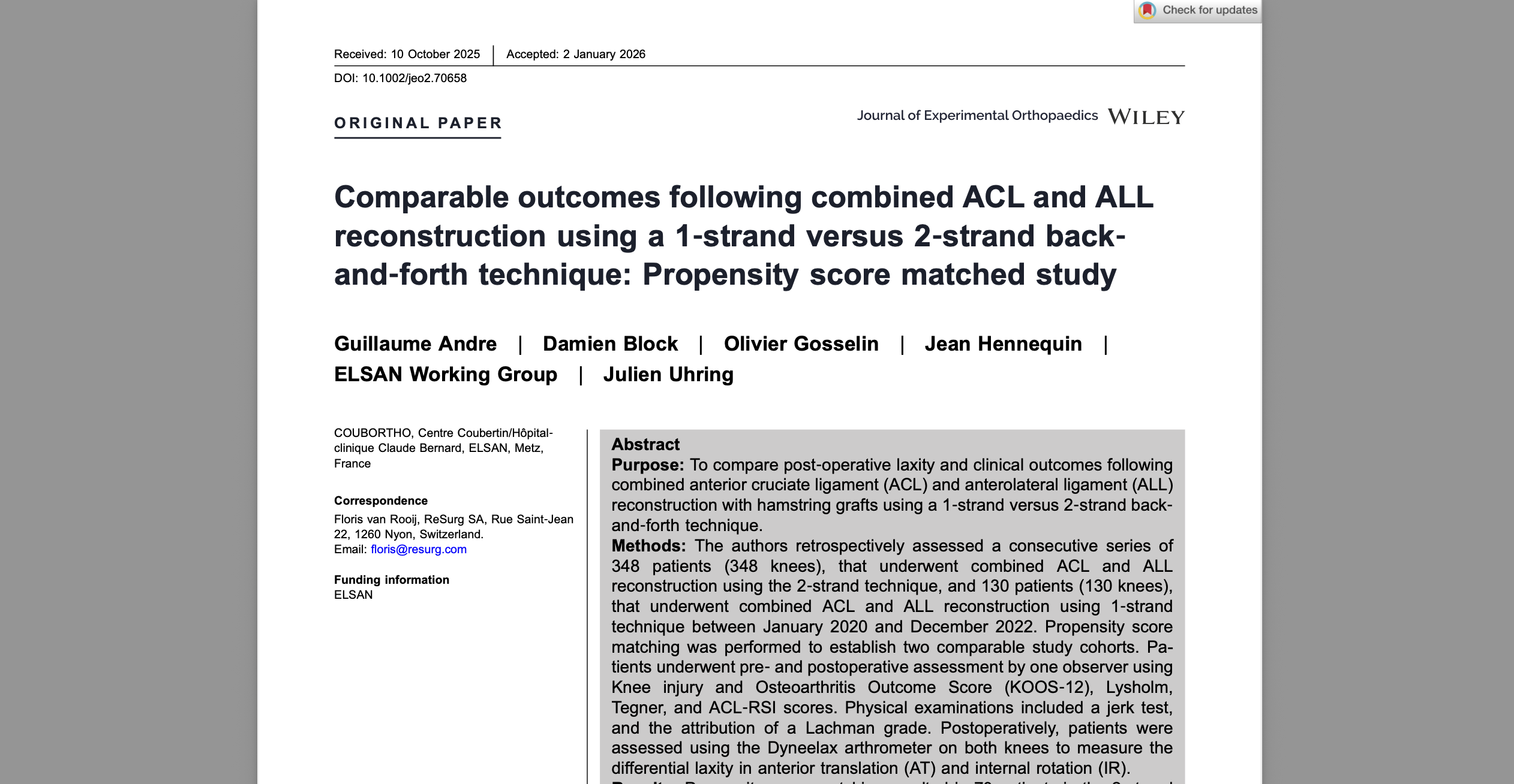
Practically, repeatable objective data can also help interpret the “partial tear” management gray zone highlighted by surgeon practice variation in Frey et al. (2025).
borderline ACL MRI instability testing is strongest when the objective number is paired with a clear narrative: what the patient feels, what the pivot shift shows (if assessable), and what MRI reveals about concomitant injury.
5) Decision aid: integrating findings into an ACL treatment decision borderline tear
This section operationalizes the workflow so that borderline ACL MRI instability testing leads to an actionable plan rather than a “watch and wait” loop.
5.1) A clinician-facing decision aid (not a guideline)
- Confirm the problem is instability: distinguish giving way from pain inhibition, patellar symptoms, or fear avoidance.
- Grade exam confidence: high (clear endpoint + consistent pivot findings) vs limited (guarding, effusion, pain).
- Cross-check MRI for modifiers: meniscal tears (ramp/root), bone contusions, collateral/PLC findings, chondral injury.
- Quantify laxity when needed: if the exam is limited or discordant, consider objective translation and document side-to-side.
- Match treatment to demand: pivoting athlete with reproducible instability is a different problem than a low-demand patient with stable exam.
- Re-test after rehab phase: if nonoperative care is chosen, reassess instability after swelling reduction and strength restoration.
This supports an ACL treatment decision borderline tear without overstating certainty: clinicians can transparently explain why the plan is rehab-first, bracing plus rehabilitation, return-to-sport criteria-based progression, or early surgical consultation.
5.2) Common pitfalls that create “borderline” confusion
- Guarding-driven false negatives on pivot shift and Lachman, especially early after injury.
- Overreliance on MRI phrasing when the patient’s instability story is consistent and reproducible, a caution echoed conceptually by Piedade et al. (2025).
- Missing a meniscal driver of instability (locking, mechanical symptoms, ramp lesions).
- Ignoring rotational contributors (anterolateral structures, PLC) when pivot shift is prominent.
5.3) Key takeaways and next steps
- borderline ACL MRI instability testing is most helpful when symptoms and MRI disagree, or when guarding limits exam reliability.
- Use a consistent sequence: history and demand profile, Lachman endpoint, pivot shift if tolerated, and evaluation of collateral/rotational contributors.
- Objective translation data can clarify equivocal cases, but it should remain complementary to MRI, which is needed to assess associated injuries and plan surgery when indicated.
- Document your correlation statement so the reasoning is clear to the multidisciplinary team and to the patient.
If uncertainty remains after a structured workup, consider a short interval reassessment after effusion control and rehabilitation, or refer for specialist opinion where imaging, exam under anesthesia, and objective measures can be integrated.
Clinical references (PubMed)
1) 2025 — Piedade et al. — Plastic deformation of anterior cruciate ligament: listen to the patient, do not just rely on imaging. — J Orthop Surg Res — DOI: 10.1186/s13018-025-05527-3 — PMID: 39915759 — PubMed
2) 2025 — Frey et al. — Management of Isolated Partial ACL Tears: A Survey of International ACL Surgeons. — Orthop J Sports Med — DOI: 10.1177/23259671241311603 — PMID: 39931635 — PubMed
3) 2025 — Khaled et al. — Complete and Partial Tears of the Anterior Cruciate Ligament: Acute and Evolution. — Semin Musculoskelet Radiol — DOI: 10.1055/s-0045-1806795 — PMID: 40393498 — PubMed
4) 2025 — Alsuwaine et al. — Chronic Bicompartmental Bucket-Handle Meniscal Tears Associated With Anterior Cruciate Ligament (ACL) Rupture in an Epileptic Patient: A Case Report. — Cureus — DOI: 10.7759/cureus.99310 — PMID: 41541969 — PubMed
5) 2025 — Choi et al. — Severe bone contusion is a risk factor for poor healing of the anterolateral ligament tear following anterior cruciate ligament reconstructions. — Knee — DOI: 10.1016/j.knee.2025.07.009 — PMID: 40743568 — PubMed