Introduction
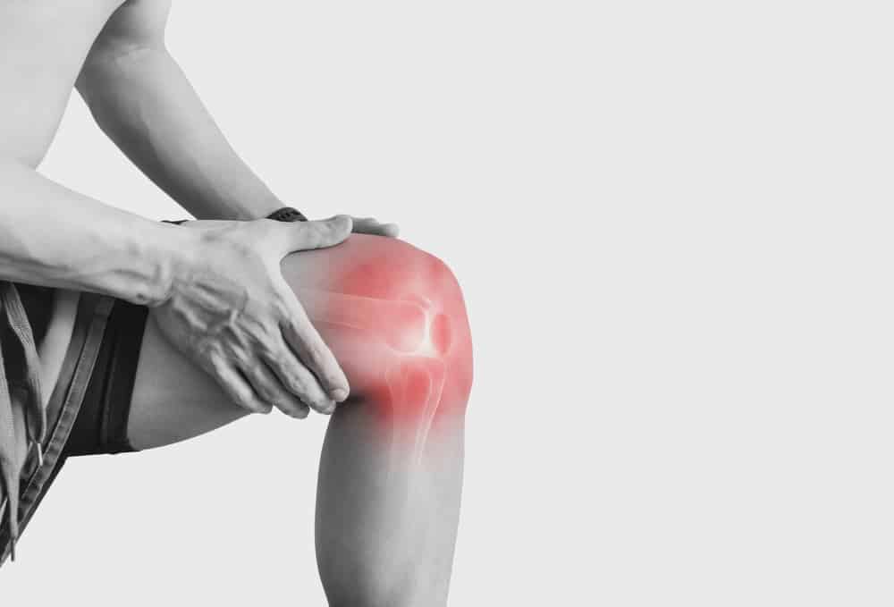
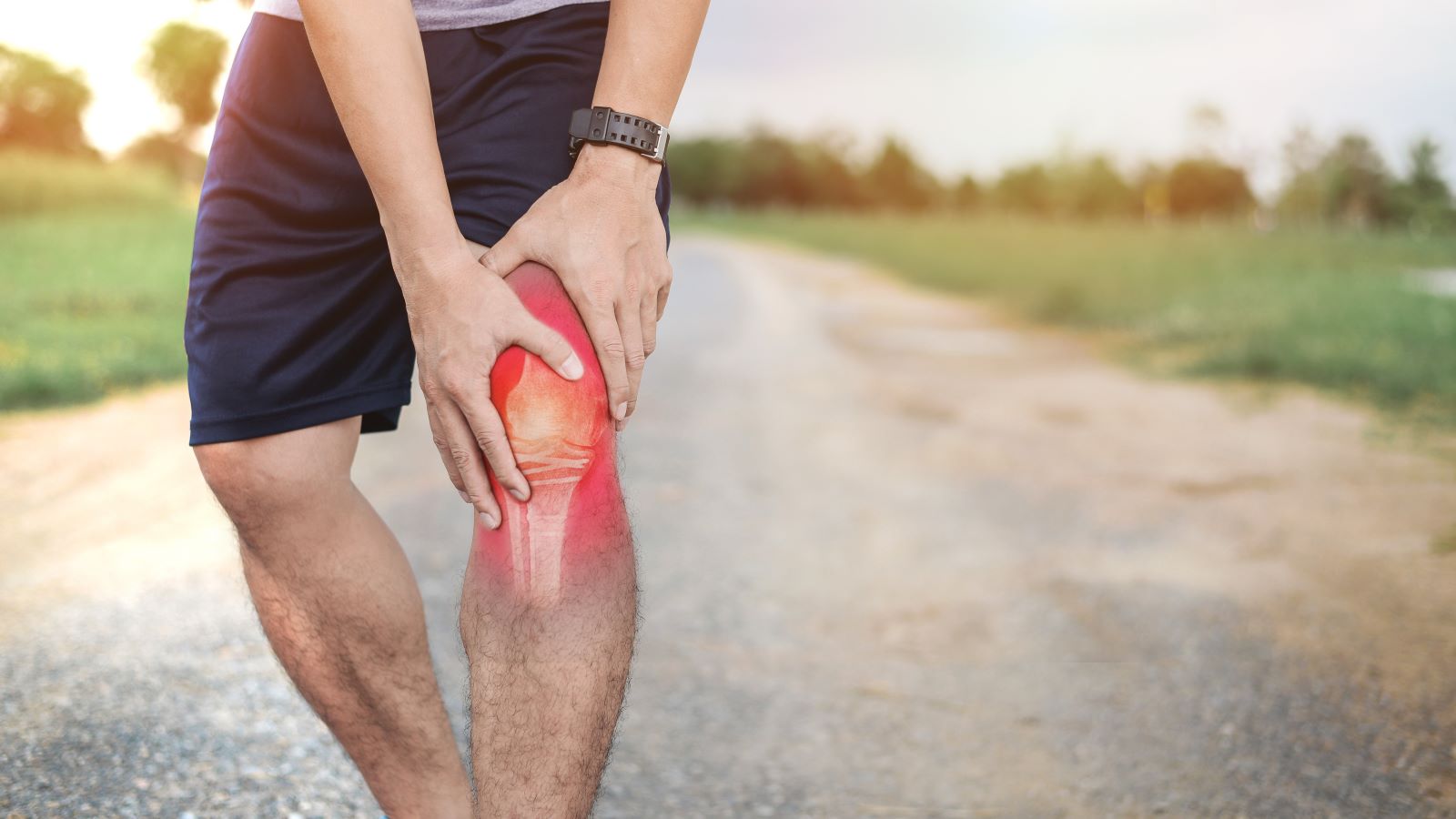
Accurate assessment of knee laxity is a cornerstone in the diagnosis and management of anterior cruciate ligament (ACL) injuries, conditions that significantly influence mobility and overall joint health. The ACL, a key ligament within the knee, is prone to injuries that can drastically affect an individual’s functional capacity. Accurate diagnosis is not just a medical priority; it’s crucial for determining the most effective treatment approach and ensuring a successful recovery. Explore Arthrometer ACL Diagnostics.
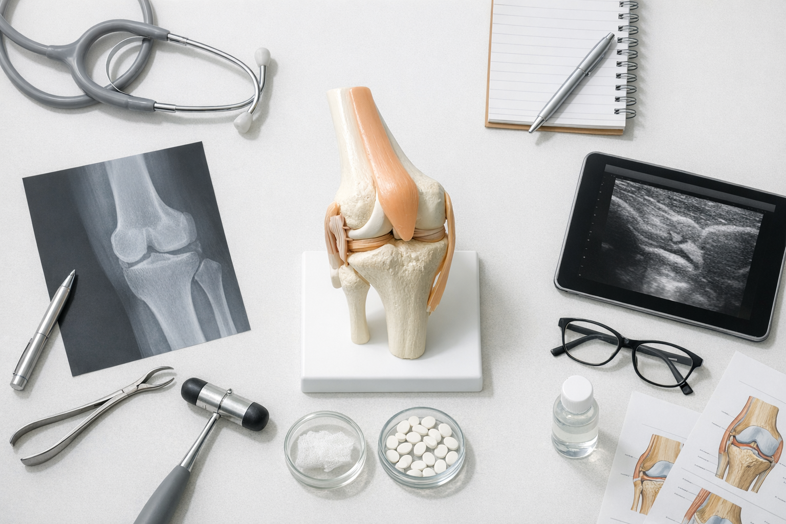
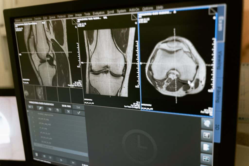
Magnetic Resonance Imaging (MRI) has traditionally been the gold standard for diagnosing ACL injuries. MRIs provide detailed visualizations of the knee’s internal structures, but they come with inherent limitations. Primarily, the static nature of MRI images does not effectively capture the dynamic stresses experienced by the knee during everyday activities. This limitation can lead to an incomplete understanding of the functional impact of ACL injuries, potentially influencing treatment decisions in a less than optimal way.
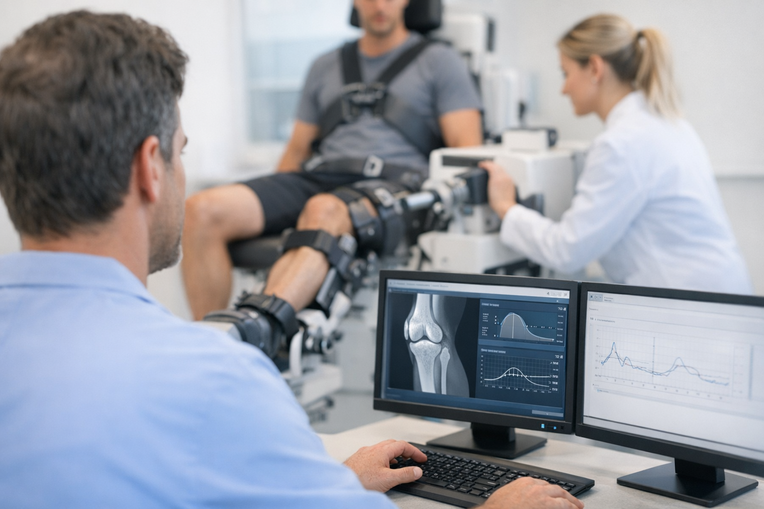
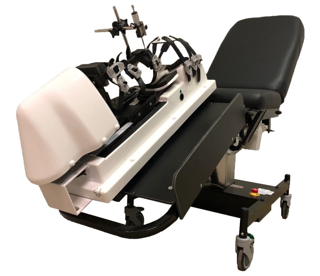
In recent years, the development of robotic arthrometers, such as DYNEELAX® and GNRB® , has introduced a novel approach to knee laxity assessment. These state-of-the-art devices represent a significant advancement, offering dynamic and functional analysis of the knee joint. Unlike MRIs, robotic arthrometers apply controlled, measurable forces to the knee, mimicking the conditions the joint faces in real-life scenarios. This approach provides a more nuanced and accurate assessment of knee stability and the condition of the ACL.
For example, the GNRB® arthrometer precisely measures knee laxity, yielding essential data in sagittal planes. On the other hand, the DYNEELAX® system evaluates translational & rotational laxity, presenting a comprehensive picture of knee stability in both sagittal and horizontal planes. Both instruments have demonstrated remarkable efficacy in diagnosing not only complete ACL ruptures but also in detecting partial tears, which are often challenging to discern via MRI.
The rise of robotic arthrometers marks a significant shift in the landscape of ACL injury diagnosis. They promise to enhance the diagnostic process, leading to more personalized treatment plans. As these technologies gain traction in clinical settings, they are poised to redefine traditional diagnostic methods, advocating for a combination of static and dynamic assessment techniques for superior patient care.
1. Current Standard: MRI in ACL Injury Diagnosis
Magnetic Resonance Imaging (MRI) remains the cornerstone in the diagnostic toolkit for anterior cruciate ligament (ACL) injuries, crucial in orthopedic and sports medicine. The ACL plays a vital role in knee stability, and injuries to this ligament can lead to substantial functional impairment. MRI’s primary role in ACL diagnosis is attributed to its capability to provide high-resolution images of the knee joint’s soft tissue structures, offering a detailed view of the ACL.
Despite MRI’s widespread use in ACL diagnostics, there are significant limitations, particularly regarding cost and the static nature of the assessment. The financial aspect of MRI is a major concern. The high cost associated with MRI scans is due to the sophisticated technology required, the expertise of specialized radiologists, and the maintenance of MRI equipment. This expense impacts healthcare systems and patients, contributing to higher overall healthcare costs. In areas where healthcare funding is limited, the cost of MRI can become a barrier to timely and adequate diagnostic care.
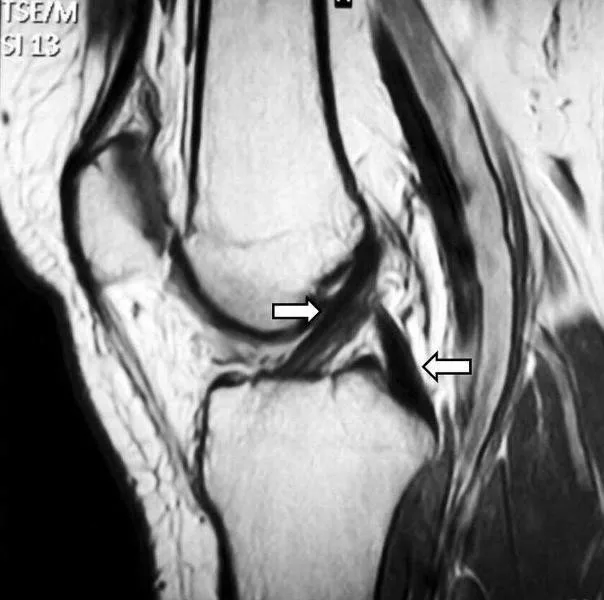
Another key limitation is the static nature of MRI assessments. ACL injuries often occur during dynamic activities, with the knee subjected to various forces and movements. However, MRI examinations are performed in a static, non-weight-bearing state, which does not mimic the typical conditions under which ACL injuries occur. This can result in a diagnostic gap, especially in evaluating the knee’s functional stability and response to stress, a vital aspect in the case of partial ACL tears or overall knee stability assessment.
Furthermore, the lack of functional assessment in static MRI images can lead to an underestimation of the injury’s severity, potentially influencing treatment decisions. For individuals with high physical demands, such as athletes, understanding the knee’s dynamic behavior post-injury is essential for effective rehabilitation and preventing recurrent injuries.
Despite these limitations, it is important to note that MRI remains an essential part of the diagnostic process when an ACL injury is confirmed. MRI is necessary to determine if other structures within the knee, such as menisci, cartilage, or other ligaments, are also damaged. While the rotational analysis provided by the Dyneelax system offers valuable insights, MRI’s comprehensive imaging is crucial for a complete understanding of the knee’s condition post-injury.
In essence, while MRI is a vital and advanced tool for diagnosing ACL injuries, its limitations in cost and static assessment underscore the need for complementary diagnostic methods. Technologies like robotic arthrometers offer dynamic, functional, and potentially more cost-effective alternatives for assessing ACL injuries, fitting into a comprehensive diagnostic approach alongside MRI.
2. The Rise of Robotic Arthrometers: GNRB and Dyneelax - Arthrometer ACL Diagnostics
In the ever-evolving field of orthopedic diagnostics, robotic arthrometers such as GNRB and Dyneelax have emerged as groundbreaking tools for the measurement of knee laxity. These devices are pivotal in advancing the diagnosis of knee injuries, especially ACL injuries, offering a depth of analysis that surpasses traditional MRI scans.
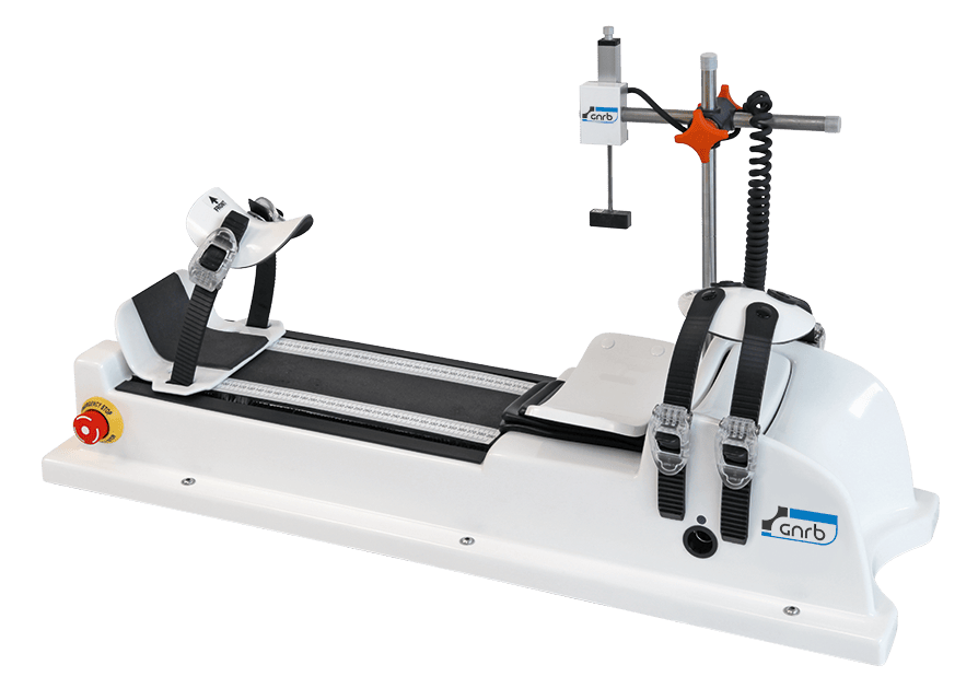
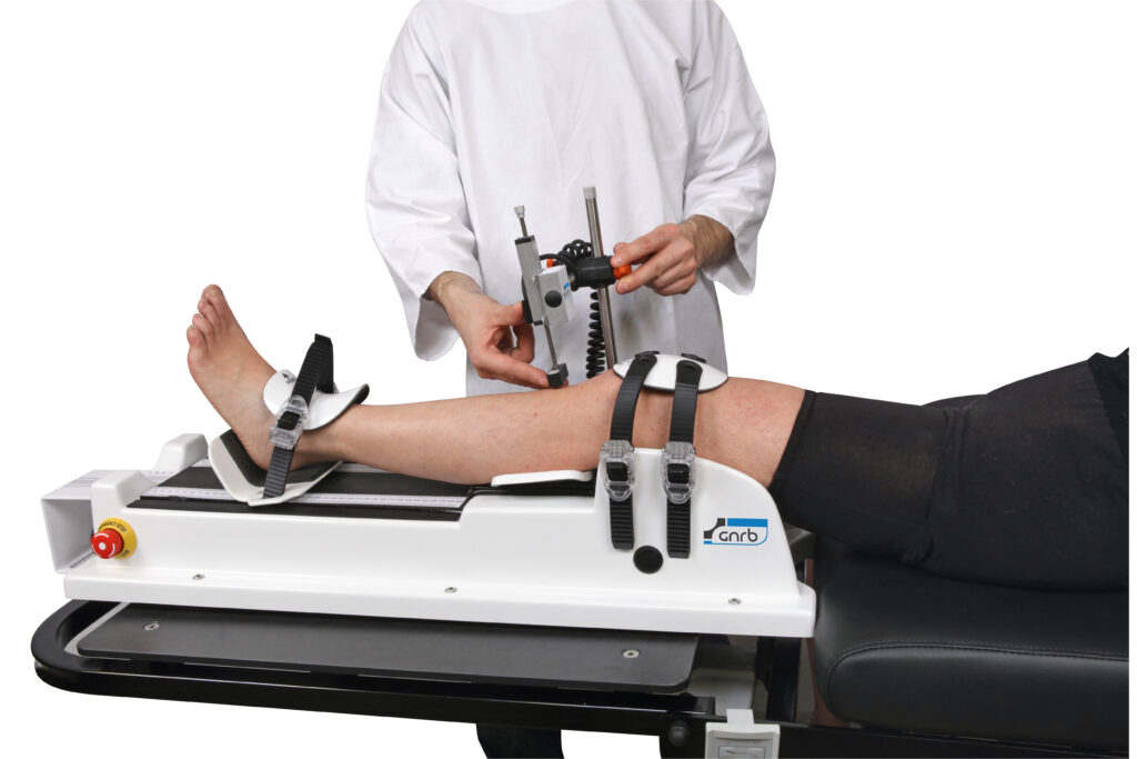
GNRB® is a technologically advanced robotic device designed to objectively assess knee laxity. It operates by applying controlled, measurable forces to the knee, effectively simulating the various stresses it encounters during daily activities and sports. GNRB® specializes in the measurement of anterior-posterior displacement of the tibia in relation to the femur, providing vital data on the stability of the ACL. Its ability to conduct assessments under dynamic conditions mirrors the real-life situations in which most knee injuries occur, offering a more authentic representation of the knee’s functional state and the extent of the injury.
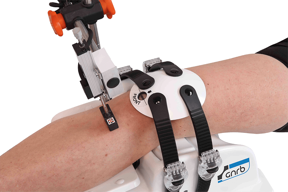
Dyneelax distinguishes itself with its ability to apply both translational and rotational forces, facilitating a comprehensive assessment of knee laxity. This dual-force application allows for a thorough examination of the knee’s stability, encompassing both translational (anterior-posterior) and rotational (lateral/medial) movements. Such a comprehensive approach is essential in accurately assessing the overall stability and functionality of the joint, particularly in sports-related injuries where both types of forces play a role. Dyneelax’s dual assessment capacity provides a critical dimension to knee diagnostics, offering a more complete understanding of knee health and potential injuries.
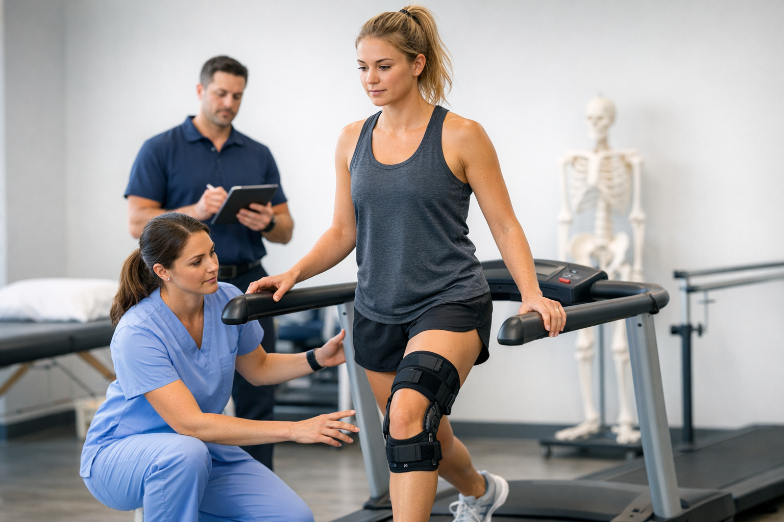
The advantages of robotic arthrometers like DYNEELAX® and GNRB® in representing real-life knee stresses are significant. Firstly, they provide a dynamic assessment of the knee, which is crucial in understanding the joint’s behavior under actual conditions. This aspect is particularly vital for athletes or individuals engaged in high-impact activities, where static assessments might not fully capture the functional implications of an injury.
Secondly, the precision and objectivity offered by these devices are unparalleled. Unlike manual testing methods, which can be subjective and vary between examiners, DYNEELAX® and GNRB® offer consistent, reproducible results. This level of objectivity is crucial in making accurate diagnoses and monitoring the progression of treatment and rehabilitation.
Additionally, these devices enhance patient engagement and understanding of their condition. Patients gain a clearer insight into the nature of their injury through the data and images provided by these arthrometers, which can be empowering in adhering to treatment plans and rehabilitation protocols.
In summary, the advent of GNRB and Dyneelax in the field of knee diagnostics marks a significant shift towards more accurate, dynamic, and comprehensive assessments of knee injuries. These technologies not only complement existing diagnostic tools like MRI but also fill critical gaps in assessing the functional impact of knee injuries. As these devices become more integrated into clinical practice, they are set to redefine the standards in knee injury diagnosis and treatment, ultimately leading to improved outcomes for patients with knee ligament injuries.
3. Clinical Efficacy (Scientific Reviews Mentioned)
The clinical efficacy and cost-effectiveness of DYNEELAX® and GNRB® arthrometers in diagnosing ACL injuries, particularly partial ruptures, have been substantiated by several significant studies, highlighting their potential in transforming current diagnostic approaches (please note that GNRB technology is incorporated in the Dyneelax).
A pivotal study by Theo Cojean in 2023 specifically addressed the efficacy of GNRB in diagnosing partial ACL ruptures. Cojean’s findings revealed that the GNRB® sensitivity and specificity were equivalent to those of MRI for healthy ACL and complete ACL tear detection. However, MRI had some difficulty in detecting partial ACL tears compared with the GNRB which showed better sensitivity. This was confirmed by arthroscopy. This indicates the GNRB’s superior capability in identifying partial ACL tears, a challenging area for conventional MRI.
Supporting the reliability of the GNRB®, Kayla Smith’s 2022 study demonstrated its consistent performance in measuring ACL stiffness and laxity. The study showed moderate to good intratester reliability, with ICC values ranging from 0.72 to 0.83, confirming the GNRB’s dependability in providing consistent assessments. Additionally, a study by Magdic in 2023 corroborated the intra-rater reliability of the GNRB, reporting good ICC values for measurements at both 134 N and 200 N forces.
While GNRB® excels in assessing anterior-posterior knee laxity, Dyneelax complements this by offering both translational and rotational laxity assessments. A key finding from another Cojean’s study on Dyneelax is that the device has excellent repeatability intra-series and good reproducibility, although it is sensitive to some positioning parameters. This dual functionality of Dyneelax is critical in providing a comprehensive assessment of knee stability, especially relevant in sports-related injuries involving rotational forces.
4. Statistical Analysis: ACL Injuries and Diagnostic Procedures
Before beginning this section, please note that our example is based on the French Healthcare System, where a robotic arthrometer test costs around 65€. It’s important to emphasize that the arguments and insights discussed can be applied globally. In our experience, the cost for arthrometer tests conducted with our devices varies internationally, ranging from 20€ to 200€.
In France, the extensive use of MRI for diagnosing knee injuries, particularly ACL ruptures, significantly influences healthcare expenditure. According to Incepto Data, approximately 1.5 million MRI tests are conducted annually for various knee issues. The “IRM du membre inférieur” document by the HAS (Haute Autorité de Santé de France) sheds further light on this, revealing that 1 out of 3 MRIs focus on the lower limb. Specifically, around 780,000 adults underwent lower limb MRI in the latter half of 2021 alone (if we multiply by two, 1.6 million french adults in 2021) , with a substantial proportion of these investigations targeting knee-related concerns.
Moreover, this document highlights a staggering 76% increase in MRI usage over the past decade. Based on these figures, it’s reasonable to estimate that around 750,000 of MRI tests each year (In 2013, Allan M. Joseph estimates ACL injuries account for half of all knee injuries; DOI: 10.4085/1062-6050-48.6.03, we will do the same) are dedicated to suspected ACL or ligament-related injuries as it is a very common knee injury.
This statistical overview underscores the heavy reliance on MRI in France for diagnosing ACL and other knee ligament injuries. It also points to a growing trend in MRI utilization, raising questions about potential overuse and the efficiency of resource allocation in the healthcare system.
While MRI remains crucial if an ACL tear is confirmed, it is interesting to put robotic arthrometers such as DYNEELAX® and GNRB® forward. However, given the discrepancy between MRI tests and actual ACL surgeries—only about 55,000 ACL reconstruction surgeries (ACLR) against an estimated 100,000 complete and partial ACL tears per year—there’s a potential overuse of MRI, with around 650,000 tests possibly being unnecessary (if there is no lesion of any ligament structures). Moreover, since tests applied by Dyneelax consist of Translation & Rotation, Translation will detect the ACL, and the rotation test will detect if other structures are also damaged. Doing a Dynamic Dyneelax test is therefore an advantage as it serves as a first filter and sends patients to do an MRI only if it is really necessary.
By adopting arthrometers for initial diagnostics, healthcare facilities can also alleviate the pressure on MRI services, currently burdened with an average wait time of 30 days (Snitem Data in 2017). This approach essentially suggests adding a crucial step in the overall treatment timeline for patients suffering from ACL injuries: Dynamic Knee Testing.
Traditionally, after an initial injury, a patient undergoes a clinical examination (like the Lachman test or the pivot shift test), followed by an MRI (a static test). If the ACL is confirmed as torn, rehabilitation or surgical intervention is considered. However, our proposed method introduces a more comprehensive approach: starting with the clinical exam (Lachman test), followed by a dynamic test replicating real-life conditions (using Dyneelax), and then, if the ACL is torn or rotational instability is detected, proceeding with an MRI (Static test). This sequential methodology—integrating clinical, dynamic, and static assessments—enables a more thorough evaluation of the injury.
Armed with this complete data set from the clinical examination, dynamic testing (Dyneelax), and static testing (MRI), healthcare professionals can make more informed decisions regarding treatment. This could range from ACL surgery alone to more complex procedures like ACL reconstruction combined with the Lemaire technique (or other antero-lateral extra articular plasty) to avoid rotational instability, or addressing multiple ligament injuries such as ACL and PCL, etc.
Incorporating this enhanced diagnostic pathway not only yields a more accurate assessment of the knee’s condition but also allows for more tailored and effective treatment plans. By integrating dynamic testing early in the diagnostic process, unnecessary MRIs can be reduced, easing wait times and resource use for MRI services. This strategy ensures that patients with ACL injuries receive comprehensive evaluations, leading to optimal treatment outcomes, while also improving the availability of MRI for patients with other urgent medical conditions, such as strokes and specific types of cancers.
To summerize, integrating robotic arthrometers into the initial diagnostic process for suspected ACL injuries offers not only enhanced diagnostic accuracy but also significant cost savings and improved management of healthcare resources. This approach promises more efficient care for various medical conditions and optimizes the use of MRI services, ensuring timely access for other critical medical conditions.
5. Financial Implications for Hospitals and Patients
Before beginning this section, please note that our example is based on the French Healthcare System, where a robotic arthrometer test costs around 65€. It’s important to emphasize that the arguments and insights discussed can be applied globally. In our experience, the cost for arthrometer tests conducted with our devices varies internationally, ranging from 20€ to 200€.
The integration of robotic arthrometers like DYNEELAX® and GNRB® into the diagnostic process for ACL injuries presents significant financial implications for both hospitals and patients in France. The acquisition cost for these arthrometers ranges between €10,000 and €50,000, which represents a considerable investment for healthcare facilities. However, this expense is offset by the substantial savings accrued from reduced MRI tests and the tests done with the arthrometer over the month following the acquisition.
For hospitals, investing in arthrometers can lead to significant long-term financial benefits. Annually, about 750,000 MRI tests are conducted in France for suspected ACL injuries, costing nearly €375 million at a rate of €500 per MRI. In contrast, utilizing arthrometers, which are priced at approximately €65 per test, could reduce this annual expenditure to around €48.75 million. This represents a potential saving for the french healthcare system of over €326 million each year. In addition, there will be less MRI tests done when there is no lesion at all (an estimated 650,000 MRI tests are done when there is no lesion of the ACL or other related ligament structures). We know that MRI allows the diagnosis of other structures but we are specifically talking about patient who are suspected to have an ACL injury in particular. Moreover, the translation test done by Dyneelax allows the analysis of the ACL while the rotation analysis allows to evaluate other structures. When results of both these tests indicate no lesion, then no MRI is needed. However, in the opposite case, MRI should be prescribed to have a more complete analysis (Dynamic + Static Testing).
Furthermore, the return on investment (ROI) for hospitals and clinics using arthrometers is compelling, especially for those that regularly encounter ACL injuries. The cost of an arthrometer can be recuperated within a few years, considering the savings from fewer MRI scans and the lower cost per arthrometer test. For example, by only doing 10 arthrometer tests per month at 65€ for ACL suspected injuries, the ROI following an initial investment of 40 000€ is 5 years. This makes arthrometers an economically viable and attractive option for healthcare providers, factoring in their added value in enhancing diagnostic accuracy and patient care.

Patients stand to benefit financially from the use of arthrometers as well. The reduced cost of arthrometer tests compared to MRIs translates into lower direct expenses, particularly in healthcare systems where patients are responsible for a portion of the diagnostic costs. This reduction in medical expenses can significantly enhance patient satisfaction, as it alleviates the financial strain associated with healthcare.
Additionally, the accuracy and timeliness of diagnoses provided by arthrometers can lead to more appropriate and effective treatment plans. The ability of arthrometers to provide dynamic testing data crucial for surgical planning can prevent unnecessary medical procedures, saving costs and reducing medical risks for patients. This not only leads to cost savings for patients but also builds trust and confidence in the healthcare system.
Reflecting on the financial impact of incorporating robotic arthrometers into the ACL injury diagnostic process reveals its substantial nature. Hospitals stand to achieve significant cost savings and improved diagnostic services, while patients are poised to benefit from reduced diagnostic costs and more accurate, timely medical care. This integration ultimately represents a sound financial strategy, crucial for enhancing both the quality and efficiency of patient care within the healthcare system.
Conclusion
In this article, we examined the impactful role of robotic arthrometers like DYNEELAX® and GNRB® in the diagnosis of ACL injuries. These advanced tools have demonstrated remarkable efficacy, especially in detecting partial ACL tears, where traditional MRIs often have limitations. Their integration into the diagnostic framework not only promises improved accuracy but also significant cost savings for healthcare systems and reduced financial burden for patients.
Looking to the future, the field of ACL injury diagnosis is on the cusp of a transformative shift. The emerging gold standard is likely to be an integrated approach that combines the precision of clinical assessments with the dynamic insights from arthrometer testing, and the comprehensive analysis provided by MRI. This multifaceted diagnostic pathway is poised to enhance overall patient care, aiming to optimize both the accuracy of diagnoses and the efficiency of healthcare delivery. This evolution in diagnostic strategies represents a significant advancement towards more personalized and effective treatment options for ACL injuries.
Medical References
- Cojean, T. (2023). MRI vs. GNRB in ACL Injuries Detection: A Comparative Study. Journal of Orthopedic Research and Therapy (DOI: 10.1016/j.knee.2023.03.017).
- Cojean, T. (2023). Sensitivity, Repeatability, and Reproducibility Study with a Leg Prototype of a Recently Developed Knee Arthrometer Dyneelax. Journal of Biomechanics (DOI: 10.1016/j.medntd.2023.100254).
- Magdic, J. (2023). Intra-rater Reliability of the Knee Arthrometer GNRB. Journal of Clinical Orthopedics (DOI: 10.1016/j.jor.2023.03.016).
- Smith, K. (2022). The Reliability of the GNRB Knee Arthrometer in Measuring ACL Stiffness and Laxity: Implications for Clinical Use and Clinical Trial Design. Journal of Orthopaedic Research (DOI: 10.26603/001c.38252)
- Joseph, A. M., Collins, C. L., Henke, N. M., Yard, E. E., Fields, S. K., Comstock, R. D. (2013). A multisport epidemiologic comparison of anterior cruciate ligament injuries in high school athletics. DOI: 10.4085/1062-6050-48.6.03