Anterior Cruciate Ligament (ACL) injuries are among the most common and debilitating knee injuries, often resulting from high-impact sports or sudden directional changes. The ACL plays a crucial role in stabilizing the knee, and its tear can lead to long-term mobility issues if not accurately diagnosed and treated promptly. Early ACL Tear Detection is vital to prevent further damage and ensure an effective recovery plan.
However, accurate detection remains a significant challenge. Traditional methods such as MRI scans and clinical examinations often fall short due to high costs, limited accessibility, and variability in results. These limitations can lead to misdiagnosis or delayed treatment, impacting patient outcomes.
At Genourob, we specialize in developing advanced knee laxity arthrometers, such as the DYNEELAX® and GNRB®. These innovative devices offer a precise, cost-effective, and reliable solution for ACL tear detection, setting a new standard in knee injury diagnosis.
I. Causes and Symptoms of ACL Tears
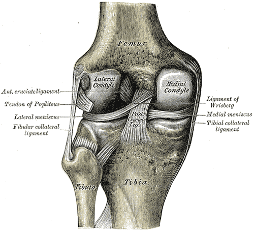
The Anterior Cruciate Ligament (ACL) is a critical ligament located in the center of the knee joint, connecting the femur (thigh bone) to the tibia (shin bone). Its primary function is to provide stability to the knee by preventing excessive forward movement of the tibia and controlling rotational forces. This stability is essential for activities involving sudden stops, jumps, or directional changes.
An ACL tear typically occurs during high-impact sports such as soccer, basketball, or skiing, where abrupt movements place excessive strain on the ligament. Common causes include landing awkwardly from a jump, pivoting sharply, or experiencing a direct blow to the knee. Symptoms of an ACL tear include a “popping” sound at the time of injury, severe pain, swelling, and instability, often leading to difficulty in bearing weight on the affected leg.
Accurate detection of an ACL tear is crucial for developing an effective treatment plan, whether it involves surgical intervention or rehabilitation. Misdiagnosis or delayed diagnosis can result in prolonged recovery, chronic knee instability, and a higher risk of further injury. Therefore, precise and timely ACL tear detection is essential for optimal recovery and returning to an active lifestyle.
II. Traditional ACL Tear Detection Methods
Traditional methods for detecting ACL tears include MRI scans, clinical examinations, manual Lachman tests, ultrasound, and manual arthrometers.
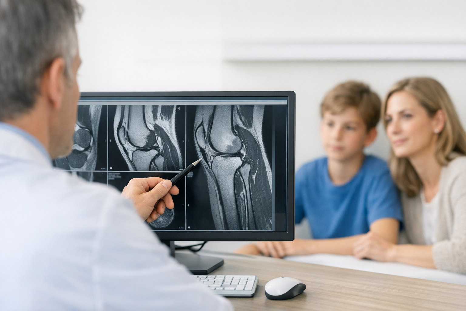
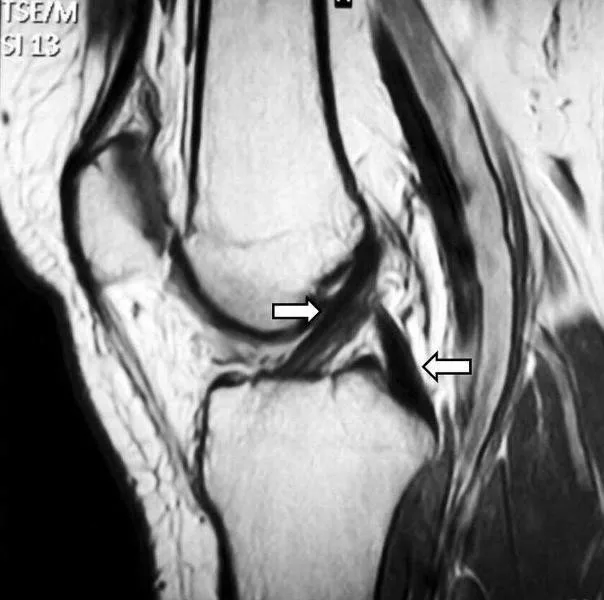
MRI (Magnetic Resonance Imaging) is a highly detailed imaging technique that visualizes the ACL and surrounding structures. While effective in identifying tears, MRI comes with significant drawbacks such as high cost, limited availability, and potential wait times for scheduling and results.
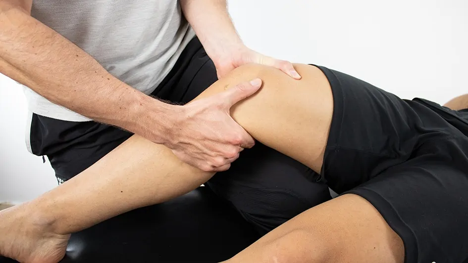
Clinical examinations, including the Lachman test and Pivot Shift test, assess knee stability through physical manipulation. These tests can provide valuable information but are highly dependent on the examiner’s skill and experience, introducing a degree of subjectivity into the diagnosis. This can sometimes lead to variability in results.
Ultrasound uses high-frequency sound waves to produce images of the knee. It is more affordable and offers real-time imaging, but may not capture the full extent of soft tissue injuries as effectively as MRI. Its effectiveness in diagnosing ACL tears can be less consistent compared to MRI.
Manual arthrometers, such as the KT-1000 & KT-2000, measure knee laxity by applying stress to the knee and assessing the degree of movement. While they provide a useful measure of knee stability, their accuracy can be influenced by the operator’s technique and the device’s calibration. Other devices such as the Telos stress Device also enable assessment of the ACL manually. The radiology nature of the test makes it however invasive. Studies have shown that manual arthrometers can have variable results, with sensitivity and specificity often dependent on the specific device and protocol used.
Overall, while these traditional methods have their applications, they each come with limitations related to cost, accessibility, and precision, which can impact the accuracy and timeliness of ACL tear diagnosis.
III. Advanced Techniques in ACL Tear Detection: GNRB and Dyneelax
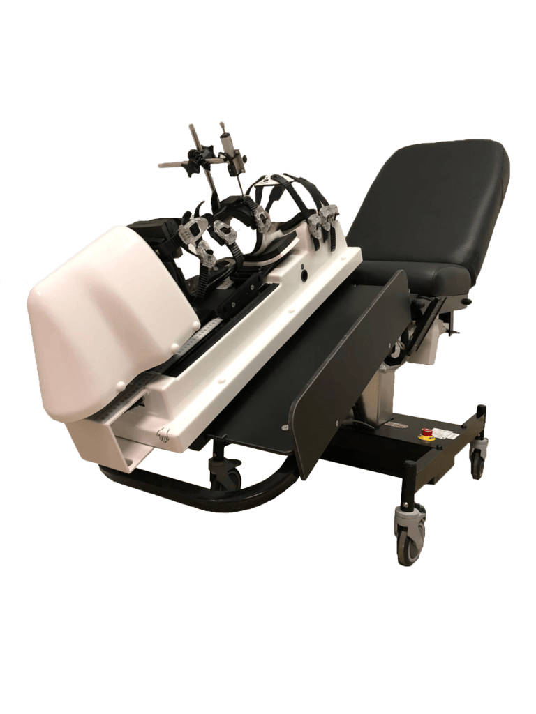
In the realm of advanced ACL tear detection, DYNEELAX® and GNRB® knee laxity arthrometers represent significant progress, offering precise and cost-effective solutions that surpass traditional diagnostic methods. These state-of-the-art devices enhance the accuracy of ACL injury assessments.
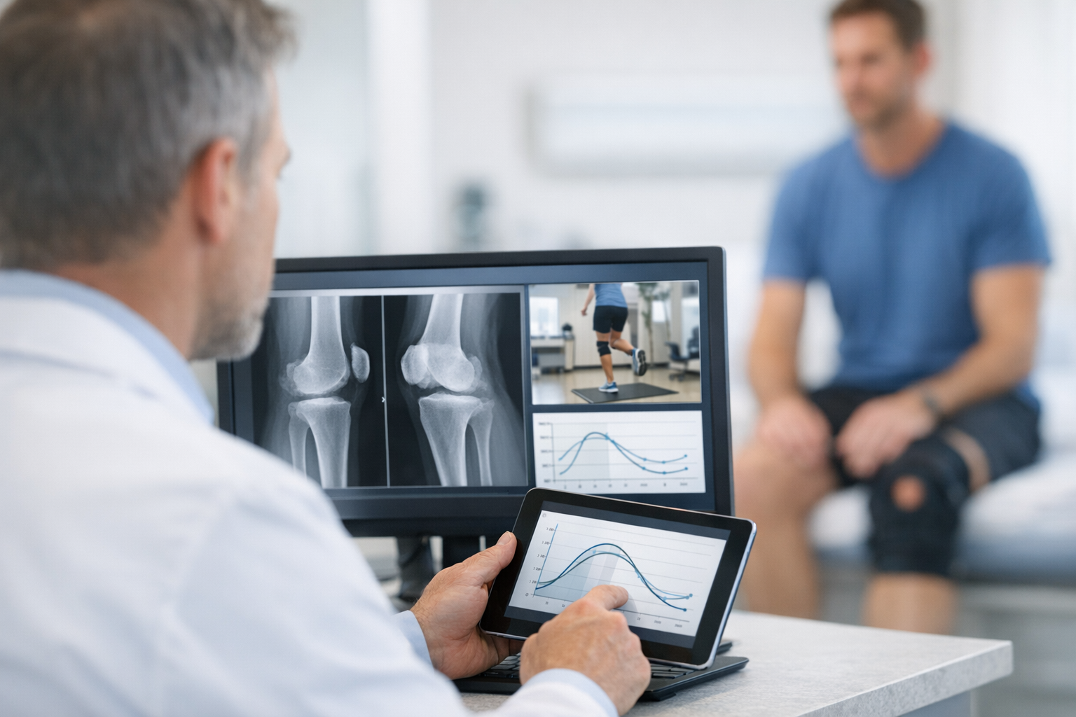
DYNEELAX® and GNRB® are specialized arthrometers designed to measure knee laxity, a crucial indicator of ACL integrity. The GNRB® applies controlled anterior tibial translation to assess knee stability. By measuring the amount of forward movement of the tibia relative to the femur, GNRB provides detailed insights into the degree of ACL damage. This method is particularly effective for evaluating the stability of the knee under stress.
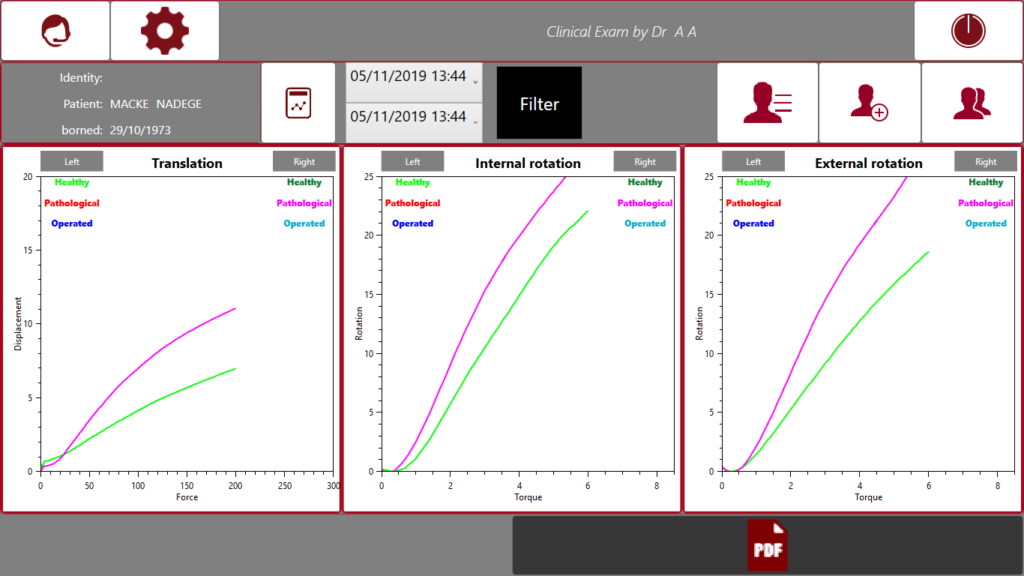
On the other hand, the DYNEELAX® device measures both anterior tibial translation and knee rotation. This dual-measurement approach offers a comprehensive assessment of knee stability, capturing both forward displacement and rotational movement. The ability to evaluate these two parameters simultaneously provides a more complete picture of ACL function and potential injury.
These devices offer several advantages over traditional methods. Their precision is a key benefit, with DYNEELAX® and GNRB® providing highly accurate measurements of knee laxity, reducing the likelihood of misdiagnosis. Their ease of use is another advantage, with intuitive interfaces and straightforward procedures that streamline the diagnostic process. Additionally, both devices are cost-effective compared to MRI, making them accessible to a wider range of healthcare settings without compromising diagnostic quality.
Compared to traditional diagnostic methods like MRI, ultrasound, and manual tests, GNRB and Dyneelax provide clear benefits. MRI, while detailed, involves high costs and long wait times. Manual tests, though useful, are subjective and reliant on the examiner’s skill, leading to variability in results. Ultrasound, though less invasive, may not capture the full extent of injuries as effectively as these advanced arthrometers.
Our robotic arthrometers, DYNEELAX® and GNRB®, are pivotal in redefining knee ligament treatment across the spectrum from injury to full recovery. They are utilised for:
- ACL Injury Diagnosis: These devices have shown superior ability in detecting partial ruptures compared to MRI.
- Surgical Planning: They enable comprehensive analysis of peripheral structures and rotational stability to determine the necessity of lateral extra-articular tenodesis (LET) surgery.
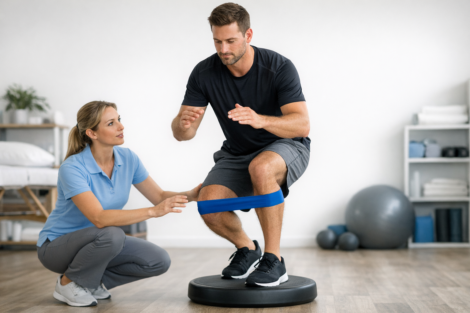
- Rehabilitation: Dyneelax plays a crucial role in personalizing rehabilitation by determining optimal return-to-sport timing and monitoring ACL grafts post-reconstruction.
These capabilities underscore the transformative impact of DYNEELAX® and GNRB® in improving diagnostic accuracy, surgical planning, and rehabilitation outcomes, establishing a new paradigm in knee ligament treatment.
IV. Case Studies
- Collette, M., Courville, J., Forton, M., & Gagnière, B. (2012). Objective evaluation of anterior knee laxity; comparison of the KT-1000 and GNRB arthrometers. Knee Surg Sports Traumatol Arthrosc. DOI: 10.1007/s00167-011-1869-2
- Lefevre, N., Bohu, Y., Naouri, J.F., Klouche, S., & Herman, S. (2013). Validity of GNRB arthrometer compared to Telos in the assessment of partial anterior cruciate ligament tears. Knee Surgery, Sports Traumatology, Arthroscopy, 22, 285-290. DOI: 10.1007/s00167-013-2384-4
- Jenny J-Y, Puliero B, Schockmel G, Harnoist S, Clavert P. (2017). Experimental validation of the GNRB® for measuring anterior tibial translation. Orthopaedics & Traumatology: Surgery & Research. Received: 7 October 2016, Accepted: 30 December 2016 DOI: 10.1016/j.otsr.2016.12.011
- Pouderoux, T., Muller, B., Robert, H. (2019). Joint laxity and graft compliance increase during the first year following ACL reconstruction with short hamstring tendon grafts.. DOI: 10.1007/s00167-019-05711-z
- Smith K, Miller N, Laslovich S. (2022). The Reliability of the GNRB® Knee Arthrometer in Measuring ACL Stiffness and Laxity: Implications for Clinical Use and Clinical Trial Design. Int J Sports Phys Ther, 17(6), 1016-1025. DOI: 10.26603/001c.38252
- Magdić M, Gošnak Dahmane R, Vauhnik R. (2023). Intra-rater reliability of the knee arthrometer GNRB® for measuring knee anterior laxity in healthy active subjects. Journal of Orthopaedics. DOI: 10.1016/j.jor.2023.03.016
- Nouveau, S., Robert, H., Viel, T. (2017). ACL Grafts Compliance During Time: Influence of Early Solicitations on the Final Stiffness of the Graft after Surgery. Journal of Orthopedic Research and Physiotherapy, 3(1), 035. DOI: 10.24966/ORP-2052/100035
- Cojean T, Batailler C, Robert H, Cheze L. (2023). GNRB® laximeter with magnetic resonance imaging in clinical practice for complete and partial anterior cruciate ligament tears detection: A prospective diagnostic study with arthroscopic validation on 214 patients. Knee, 42, 373-381. DOI: 10.1016/j.knee.2023.03.017
- Forelli F, Le Coroller N, Gaspar M, et al. (2023). Ecological and Specific Evidence-Based Safe Return To Play After Anterior Cruciate Ligament Reconstruction In Soccer Players: A New International Paradigm. IJSPT. Published online April 2, 2023. DOI: 10.26603/001c.73031
- Cojean, T., Batailler, C., Robert, H., Cheze, L. (2023). Sensitivity repeatability and reproducibility study with a leg prototype of a recently developed knee arthrometer: The DYNEELAX®. Medicine in Novel Technology and Devices, 19, 100254. DOI: 10.1016/j.medntd.2023.100254
- Guegan, T. (2024). All the menisco‐ligamentary structures of the medial plane play a significant role in controlling anterior tibial translation and tibial rotation of the knee. Cadaveric study of 29 knees with the Dyneelax® laximeter. DOI: 10.1002/jeo2.12038
- Nascimento, N., Kotsifaki, R., Papakostas, E., Zikria, B. A., Alkhelaif, K., Hagert, E., Olory, B., D’Hooghe, P., & Whiteley, R. (2024). DYNEELAX Robotic Arthrometer Reliability and Feasibility on Healthy and Anterior Cruciate Ligament Injured/Reconstructed Persons. Translational Sports Medicine, 2024(1), 1-5. DOI: 10.1155/2024/3413466
Conclusion
In summary, ACL tear detection is a critical aspect of effective knee injury management, and traditional methods like MRI and manual tests, while useful, have limitations in precision, cost, and accessibility. GNRB and Dyneelax represent a significant advancement in knee laxity measurement, offering unparalleled accuracy in detecting both complete and partial ACL tears. These devices not only enhance diagnostic precision but also support surgical planning and personalized rehabilitation, making them indispensable tools in modern orthopedic practice. Healthcare professionals are encouraged to adopt these advanced techniques to provide their patients with more accurate, cost-effective diagnoses and treatment pathways.