In acute medial-sided knee pain, the MCL valgus stress test remains a fast, high-yield maneuver to guide triage, imaging, and early bracing decisions, especially when swelling and apprehension limit more complex exams. This workflow focuses on reproducible positioning, interpretation, and documentation so you can translate “opens up a bit” into clinically actionable language such as medial joint line gapping patterns, end point quality, and side-to-side asymmetry. It also connects bedside findings to MCL injury grading, clarifies when MRI is complementary (meniscus, cartilage, bone bruise, multiligament planning), and outlines follow-up strategies for return-to-sport and work decisions in orthopaedics, sports medicine, physiotherapy, and the ED.
1. Workflow entry: history, red flags, and medial complex map
Start with mechanism and function. A valgus force with external rotation (contact or non-contact) raises suspicion for a medial collateral ligament sprain and possible anteromedial rotatory involvement. Ask about a “pop,” immediate swelling (hemarthrosis suggests intra-articular injury), subjective giving way, and whether the patient could continue activity.
Safety first (especially in the ED). Before you perform the MCL valgus stress test, screen for fracture (tibial plateau, femoral condyle), gross dislocation history, neurovascular compromise, and severe pain that suggests the need for analgesia, immobilization, or urgent imaging. A quick comparison to the contralateral side helps contextualize baseline what is knee laxity in that patient (constitutional laxity, prior injury, or surgery).
1.1. Medial stabilizers you are actually testing
Valgus stability is shared across the superficial MCL (sMCL), deep MCL, posterior oblique ligament (POL), capsule, and secondary restraints including cruciates and the posteromedial corner. Keeping this anatomy in mind improves interpretation and referral decisions. A practical review of medial plane knee structures involved in valgus stability can help align your exam wording with what imaging and surgical colleagues expect.
Clinical note on workflow consistency. If your service is trying to standardize documentation across clinicians, embed valgus findings into a broader objective knee examination in orthopaedics template so side-to-side comparisons are captured systematically (pain, gapping, end point, and rotational coupling).
2. Performing the MCL valgus stress test: setup to endpoint
The goal is a repeatable, patient-tolerant maneuver that separates pain inhibition from true laxity. The maneuver is sometimes described generically as the valgus stress test knee, but consistency in angles and stabilization is what makes results comparable between clinicians and over time.
2.1. Patient positioning and hand placement (reduce guarding)
- Position: Supine. Expose both knees for side-to-side comparison.
- Hip: Slight abduction and external rotation can relax adductors if the patient is guarding.
- Stabilize the femur: One hand on the lateral distal femur (or thigh against your torso) to limit hip motion.
- Control the tibia: Other hand at the medial distal tibia/ankle to apply valgus while maintaining neutral tibial rotation unless you are deliberately assessing rotational coupling.
2.2. Two-angle testing (why 30 degrees and 0 degrees matter)
At about 30 degrees flexion: the MCL valgus stress test is typically most sensitive for sMCL insufficiency because bony congruence is reduced and cruciate contribution is less dominant. Compare the amount of opening and the end point quality to the contralateral knee.
At 0 degrees (full extension): repeat the MCL valgus stress test to assess for combined injury patterns. Increased valgus opening in extension may raise concern for POL/capsular injury, cruciate involvement, or more global medial complex disruption. Pain at extension can also reflect meniscal pathology or an osteochondral lesion, so interpret cautiously.
2.3. Documentation that supports decisions
Document the MCL valgus stress test with language that other clinicians can act on:
- Pain: none, mild, moderate, severe (and where).
- Gapping: none, mild, moderate, marked, plus a side-to-side impression.
- End point: firm, soft, absent.
- Angle tested: 30 degrees and/or 0 degrees.
- Confounders: effusion, guarding, analgesia, prior contralateral laxity.
2.4. Common pitfalls that create false positives or false negatives
Pain inhibition can mimic instability, while strong hamstring co-contraction can mask it. Other pitfalls include unrecognized tibial external rotation (can exaggerate medial opening), inadequate femoral stabilization, and failing to compare to the other side.
3. Interpretation and MCL injury grading (AM/PM and POL)
Your exam has two outputs: (1) a clinical estimate of injury severity and (2) an initial hypothesis of which structures are involved (anterior vs posterior medial complex, plus rotational features). In many settings, the first actionable step is determining whether functional valgus restraint is preserved, because this influences bracing, weight-bearing, and referral urgency.
3.1. Practical MCL injury grading from bedside findings
For MCL injury grading, anchor your interpretation to the MCL valgus stress test using three clinician-friendly descriptors: pain pattern, amount of gapping (relative to the other knee), and end point quality.
- Low-grade sprain (often Grade I): pain with a firm end point and little to no side-to-side gapping.
- Partial tear (often Grade II): more noticeable opening with a softer end point compared with the contralateral knee.
- Complete tear (often Grade III): marked opening with a poor or absent end point; consider associated injuries and posteromedial corner involvement.
Because baseline laxity varies, describe findings as “side-to-side increased opening” rather than relying on a single absolute distance unless you have quantified imaging. The conceptual frame of ligamentous laxity is useful here: laxity is a functional property influenced by tissue integrity, neuromuscular control, and patient-specific baseline.
3.2. AM vs PM cues (why the “medial complex” matters)
If valgus laxity couples with external rotation or anteromedial rotatory symptoms, consider broader medial-sided injury patterns rather than an isolated sMCL problem. When the MCL valgus stress test suggests Grade III behavior, consider whether the POL and posteromedial capsule are also compromised, especially if extension testing is abnormal or the patient reports instability in terminal extension.
For clinicians aligning exam impressions with operative planning discussions, the reconstruction literature is evolving. For example, Ade-Conde et al. (2026) reviews outcomes for combined MCL and POL reconstruction in Grade III injuries, supporting the concept that addressing both structures may help restore medial stability in selected patients. Technique selection remains surgeon-led and context-dependent, and conservative management remains appropriate for many isolated injuries.
3.3. Combined ligament context (do not overinterpret a single test)
In combined ACL and medial-sided injuries, clinical laxity patterns can be altered by effusion, guarding, and anterior translation. In these cases, repeat the MCL valgus stress test after aspiration (if indicated and within local practice), analgesia, or short-interval re-examination can improve confidence.
For combined ACL and MCL pathways, graft and timing decisions depend on instability pattern, sport demands, and associated pathology. Evidence syntheses such as Touhey et al. (2025) and comparative clinical discussions like Chung et al. (2024) can inform team conversations, but they do not replace individualized decision-making based on exam, MRI, and patient goals.
4. When findings are equivocal: imaging, quantification, and differential diagnosis
Equivocal bedside exams are common in the first 72 hours due to pain and swelling. If the MCL valgus stress test is limited by guarding, consider a short, structured plan: protect the knee, control effusion, reassess, and escalate imaging based on red flags and functional instability.
4.1. MRI and ultrasound positioning (complementary, not competing)
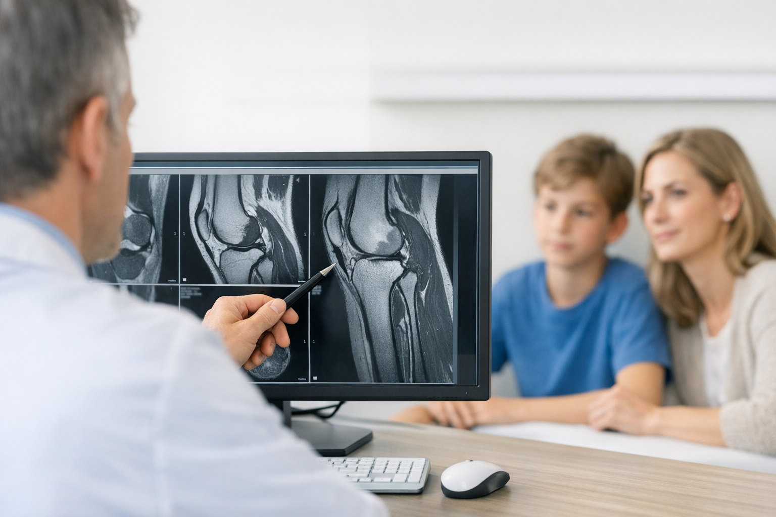
MRI is complementary to clinical testing because it evaluates associated injuries (meniscus, cartilage, bone bruise, cruciates) and helps pre-operative planning when reconstruction is considered. Ultrasound can support dynamic assessment of the superficial MCL in experienced hands, but operator dependence is a limitation.
4.2. Quantifying medial opening: stress radiography and device-based approaches
If your pathway requires objective quantification, stress radiography valgus can document medial compartment opening under standardized load. A cadaveric robotic study, Briese et al. (2025), supports that stress radiography measures correlate with severity of medial-sided injury and can be reliable in controlled conditions. In practice, technique standardization (positioning, force application, and consistent landmarks) is critical.
When considering stress methods, be explicit in documentation about the device and setup used, because values are not directly interchangeable across systems. A practical starting point is understanding the pros and cons outlined in GNRB and Dyneelax vs Telos stress device, particularly around standardization and repeatability in follow-up.
4.3. Decision aid for escalation (clinic and ED)
- Confirm stability in extension: abnormal opening at 0 degrees increases concern for combined injury.
- Check for associated injuries: locking, large effusion, inability to bear weight, or neurovascular symptoms warrant urgent imaging or referral.
- Decide on immobilization vs hinged bracing: based on pain, control, and suspected grade.
- Plan a reassessment window: early re-exam (often within days) can clarify laxity once pain improves.
- Order MRI when indicated: especially with suspected multiligament injury, meniscal symptoms, or persistent functional instability.
4.4. Differential diagnosis that mimics medial laxity
Your differential diagnosis MCL tear should revisit the MCL valgus stress test findings in light of alternative or coexisting problems: medial meniscus tear, osteochondral injury, tibial plateau fracture, pes anserine pathology, posteromedial corner injury, or referred pain from hip/adductor. If anteromedial rotatory instability is suspected, adjunctive procedures have been described in selected cases; for example, Azuma et al. (2026) reports semimembranosus transposition as an augmentation technique in a retrospective series, which is not a substitute for careful diagnosis and standard reconstruction principles.
5. Monitoring recovery and return-to-sport decisions
In most isolated injuries, the near-term goal is restoring comfort and confidence while protecting healing tissue. MCL rehabilitation monitoring should be structured so that symptoms, function, and stability are tracked in a way that can change the plan when progress stalls.
5.1. What to re-check at each follow-up
MCL rehabilitation monitoring starts with repeating the MCL valgus stress test at the same angles and with the same stabilization each time, ideally by the same clinician or with standardized documentation. Pair this with:
- Effusion and pain: trend rather than single timepoints.
- ROM milestones: extension symmetry and flexion tolerance.
- Gait quality: valgus collapse, apprehension, and brace reliance.
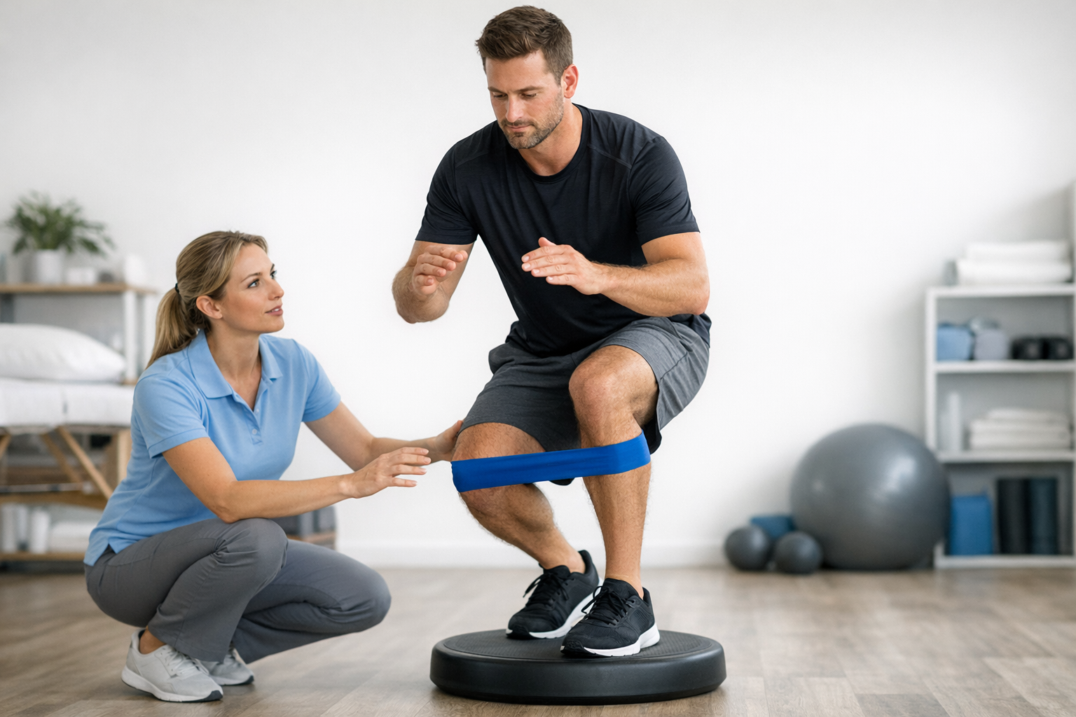
- Functional tasks: step-down control, single-leg stance, sport-specific change-of-direction progression.
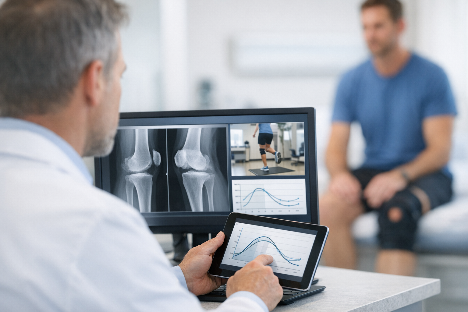
At follow-up, use the MCL valgus stress test alongside functional hop testing and movement-quality assessments to avoid clearing athletes based on symptoms alone. Where available, adding standardized objective measures can improve communication across the team and help justify progression or delay.
5.2. Where objective laxity measurement can fit (optional, pathway-dependent)
For clinics that track knee valgus laxity measurement over time, instrumented laxity testing can complement the manual exam by standardizing load application and quantifying side-to-side differences, alongside MRI when internal derangement is suspected. Depending on local availability and indications, systems used in broader laxity assessment may include the GNRB arthrometer for objective knee laxity testing and the Dyneelax device for dynamic knee laxity evaluation, with results interpreted in clinical context rather than as stand-alone diagnoses.
If you are building a measurement pathway, align it with an overview of knee laxity testing methods and consider how your documentation integrates with dynamic knee testing when rotational symptoms persist.
5.3. Documentation that supports shared decisions
In addition to narrative notes, consider a simple longitudinal table in the chart that includes pain score range, effusion grade, ROM, brace status, and a standardized statement of medial opening. For teams using objective follow-up tools, resources on objective knee joint laxity evaluation for follow-up and an orthopaedic testing device for knee ligament assessment can help with consistent implementation.
Surgical context (selected patients). Persistent symptomatic medial instability, multiligament injury, bony avulsion, or high-demand athletes with combined injuries may progress to operative discussion. When surgical approaches are debated, comparisons such as Lind vs LaPrade medial reconstruction can help frame expectations about controlling translation and rotation, but the final plan remains clinician-led, anatomy-specific, and imaging-informed.
6. Key takeaways and next steps
Use the same angles, the same stabilization, and the same words. Reproducibility is what turns a bedside exam into a longitudinal clinical tool.
- The MCL valgus stress test at ~30 degrees mainly informs sMCL integrity; abnormal opening in full extension increases concern for combined injury.
- Translate findings into decisions: early protection, targeted imaging, and planned re-exam when pain limits reliability.
- If the MCL valgus stress test remains asymmetric with a soft end point after appropriate rehab and time, reconsider the diagnosis (posteromedial corner, cruciate involvement, meniscus) and escalate assessment with MRI and/or standardized stress measurement where appropriate.
Clinical references (PubMed)
1) 2025 – Briese et al. – Stress radiography of medial knee instability provides a reliable correlation with the severity of injury and medial joint space opening-A robotic biomechanical cadaveric study. – Knee Surgery, Sports Traumatology, Arthroscopy – DOI: 10.1002/ksa.12594 – PMID: 39838900 – PubMed
2) 2024 – Chung et al. – Effect of Graft Choice for ACL Reconstruction on Clinical Outcomes in Combined ACL and MCL Injuries: Comparison Between Bone-Patellar Tendon-Bone and Hamstring Autografts. – J Clin Med – DOI: 10.3390/jcm13216316 – PMID: 39518456 – PubMed
3) 2026 – Ade-Conde et al. – Combined medial collateral ligament and posterior oblique ligament reconstruction demonstrates favourable patient-reported outcomes and medial knee stability in Grade III injuries: A systematic review. – Knee Surgery, Sports Traumatology, Arthroscopy – DOI: 10.1002/ksa.70344 – PMID: 41711578 – PubMed
4) 2026 – Azuma et al. – Transposition of the Semimembranosus as an Augmentation Technique for Anteromedial Rotatory Instability of the Knee: A Retrospective Case Series Study. – J Knee Surg – DOI: 10.1055/a-2779-0226 – PMID: 41529724 – PubMed
5) 2025 – Touhey et al. – Concomitant Anterior Cruciate Ligament and Medial Collateral Ligament Reconstruction: A Systematic Review. – Orthop J Sports Med – DOI: 10.1177/23259671251369019 – PMID: 40980559 – PubMed