Enhancing ACL Treatment from Injury to Recovery with Robotic Arthrometers
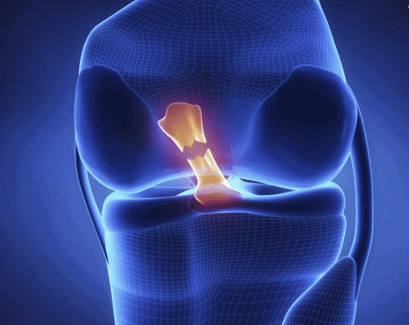
Anterior Cruciate Ligament (ACL) injuries pose a significant challenge for both patients and healthcare providers. Traditional diagnostic and rehabilitation tools have offered limited insights into the complex biomechanical properties of the knee, often resulting in suboptimal outcomes. However, recent advancements with robotic arthrometers, particularly the DYNEELAX® and GNRB® systems, have revolutionized the landscape of ACL management. These devices are not merely replacements for manual arthrometers; they are transformative tools that enhance every stage of ACL treatment—from injury diagnosis to full recovery.
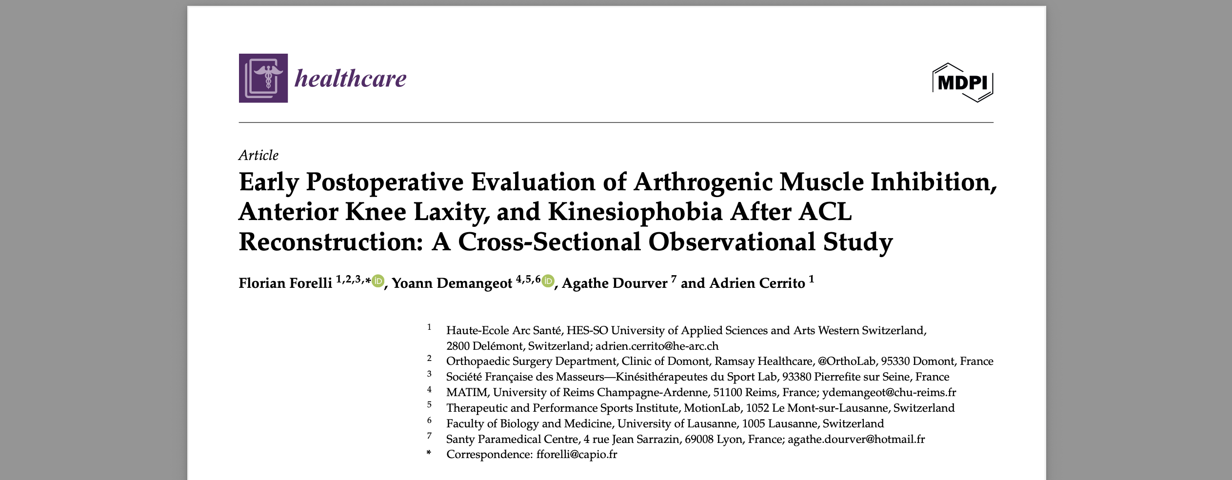
This article delves into three pivotal studies that highlight the superiority of robotic arthrometers and their role in redefining ACL treatment protocols.
I. Dynamic ACL Testing with DYNEELAX® & GNRB® Outperforms MRI for Partial Tears
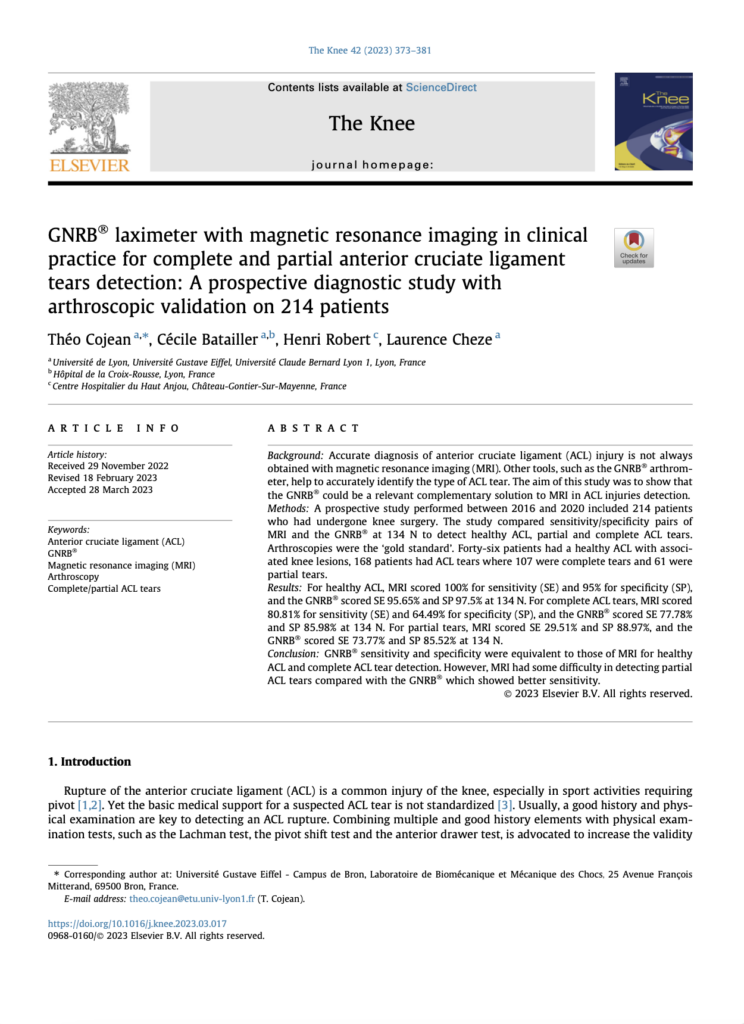
The study by Cojean et al. (2023) compared the diagnostic performance of the GNRB® robotic arthrometer with Magnetic Resonance Imaging (MRI) in detecting partial and complete ACL tears. The prospective analysis included 214 patients who underwent arthroscopic validation—the gold standard for ACL tear assessment. Key findings revealed:
Partial ACL Tears: MRI demonstrated only 29.51% sensitivity, whereas the GNRB® achieved an impressive 73.77% sensitivity at 134N force application. This highlights the GNRB®‘s ability to identify subtle changes in knee laxity, which are often overlooked by imaging techniques.
Complete ACL Tears: Both tools showed comparable sensitivity; however, the GNRB® offered superior specificity (85.98%) compared to MRI (64.49%).
The implications are profound. Dynamic laxity testing with GNRB® allows clinicians to differentiate between partial and complete ACL tears more accurately. This is particularly crucial when tailoring surgical or conservative treatment strategies. Unlike static imaging, the GNRB® provides quantitative, reproducible data on anterior tibial translation, enhancing the precision of clinical decision-making.
Reference: Cojean, T. et al. (2023). DOI: 10.1016/j.knee.2023.03.017
II. Guiding Surgical Strategies with Dyneelax®
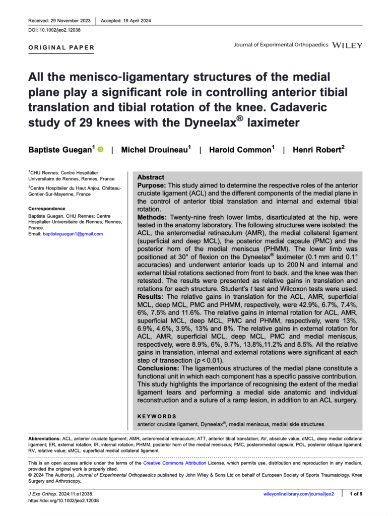
Baptiste Guegan et al. (2024) conducted a groundbreaking cadaveric study utilizing the DYNEELAX® laximeter to assess the contributions of medial knee structures to anterior tibial translation (ATT) and rotational stability. The study evaluated 29 knees and systematically sectioned key ligaments, including the ACL, anteromedial retinaculum (AMR), and medial collateral ligaments (MCL).
Key findings included:
The ACL accounted for 42.9% of anterior translation restraint, confirming its primary role in sagittal stability.
Secondary structures such as the posterior horn of the medial meniscus (PHMM) and the posteromedial capsule (PMC) contributed 11.6% and 7.5% of restraint, respectively.
Internal and external rotational gains after ligament transections underscored the functional interplay of these structures.
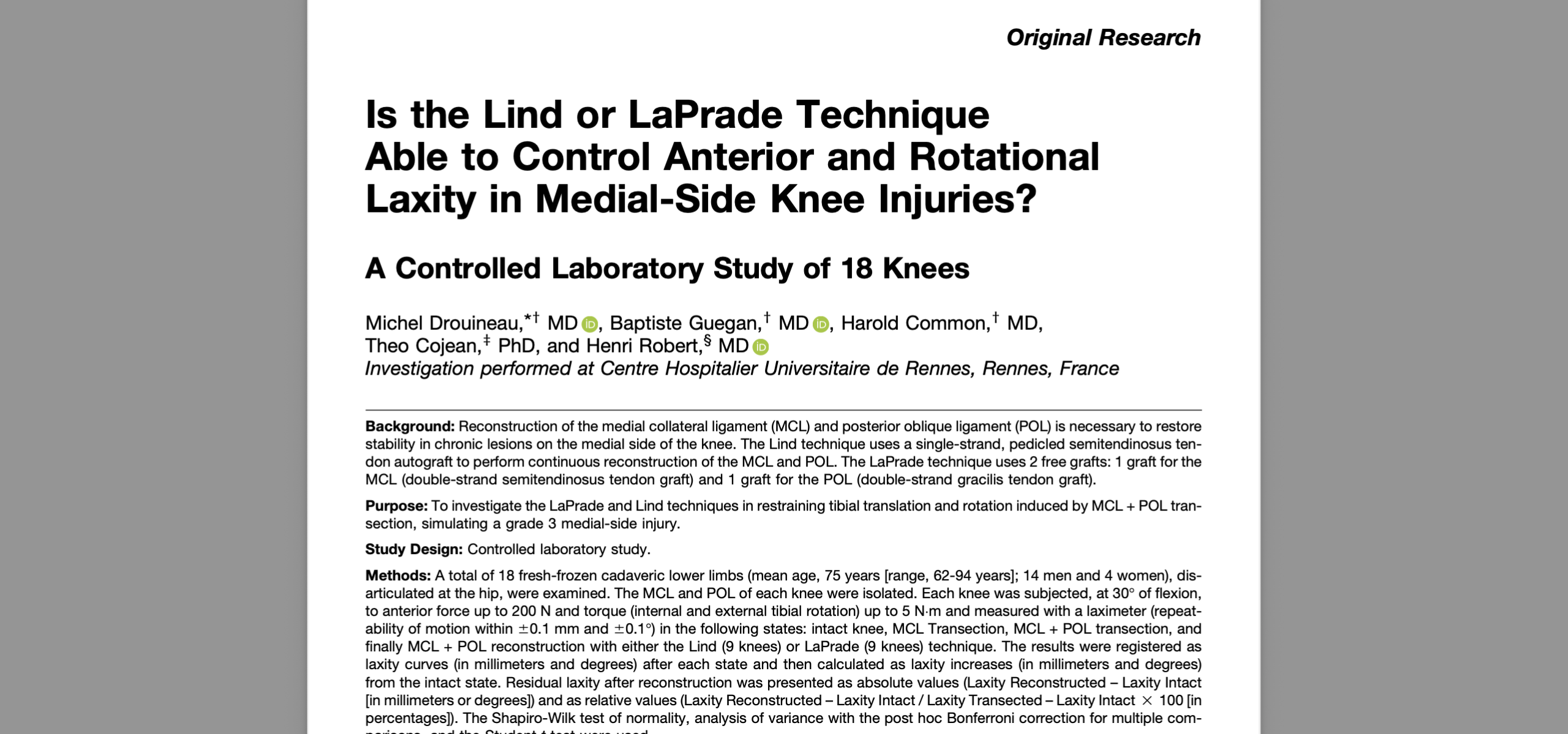
This study demonstrates how robotic arthrometers like DYNEELAX® enable precise evaluation of ligamentous contributions to knee stability. Such data guides surgeons in deciding whether isolated ACL reconstruction suffices or if additional procedures (e.g., lateral extra-articular tenodesis [LET] or medial reconstruction techniques) are required. By incorporating dynamic rotational and translational measurements, Dyneelax® ensures a more comprehensive understanding of multiligament injuries.
Reference: Guegan, B. et al. (2024). DOI: 10.1002/jeo2.12038
III. Personalizing Rehabilitation with DYNEELAX ® & GNRB®
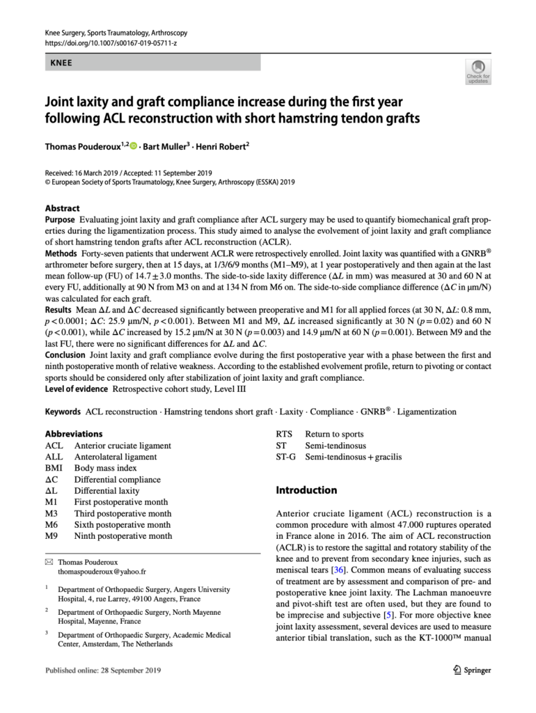
Thomas Pouderoux et al. (2019) explored how robotic arthrometers can monitor graft compliance and knee laxity during the postoperative phase of ACL reconstruction. The study involved 47 patients who underwent regular assessments with the GNRB®over 14 months.
Key observations included:
Early Stabilization: Significant reductions in joint laxity and graft compliance were observed within the first month post-surgery, indicative of effective initial graft fixation.
Ligamentization Phase: Between months 1 and 9, laxity and compliance increased slightly, reflecting the remodeling process of graft maturation.
Late Phase Stability: After nine months, joint stability plateaued, suggesting readiness for return-to-sport assessments.
These findings underscore the importance of dynamic knee laxity monitoring to tailor rehabilitation protocols. The GNRB® enables clinicians to detect phases of graft vulnerability and adjust exercises or activity levels accordingly, minimizing the risk of re-injury. Unlike manual devices, the GNRB®’s high reproducibility ensures consistent tracking of biomechanical changes over time.
Reference: Pouderoux, T. et al. (2019). DOI: *10.1007/s00167-019-05711-z
IV. The Added Value of Robotic Arthrometers
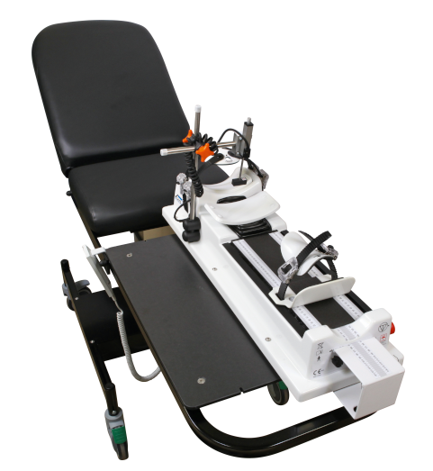
Robotic arthrometers, particularly the DYNEELAX® and GNRB®, represent a significant leap forward in orthopedic diagnostics and treatment, offering unparalleled advantages over traditional manual devices. Unlike manual arthrometers, which are limited by operator variability and lack of dynamic data, robotic systems provide precise, reproducible, and comprehensive insights into knee stability.
-
Enhanced Precision: Automated force application and displacement measurement eliminate operator variability, ensuring reproducibility. This precision is critical for accurate diagnosis and monitoring, particularly in complex cases like partial ACL tears.
-
Comprehensive Diagnostics: Dynamic testing captures both sagittal and rotational laxity, offering a holistic view of knee stability. By measuring both anterior tibial translation and rotational parameters, robotic arthrometers provide insights that manual devices cannot match.
-
Clinical Versatility: From initial diagnosis to postoperative monitoring, robotic arthrometers support informed decisions throughout the treatment journey. Their ability to assess the knee’s biomechanical properties over time ensures that clinicians can adapt treatment protocols to the patient’s evolving needs.
-
Surgical Planning: Insights into ligamentous contributions guide tailored reconstruction techniques. For example, the Dyneelax® can differentiate the roles of secondary stabilizers like the medial meniscus and posteromedial capsule, helping surgeons decide whether additional procedures like LET or MET are required.
-
Personalized Rehabilitation: Real-time data on graft behavior enable customized protocols, optimizing recovery outcomes. Continuous monitoring ensures that patients can safely progress through rehabilitation without risking re-injury.
By integrating these advanced capabilities, robotic arthrometers redefine the standard of care in ACL treatment, empowering clinicians to deliver superior outcomes and improving the overall patient experience.
V. Business Model and Economic Impact of Robotic Arthrometers
Robotic arthrometers, such as the DYNEELAX® and GNRB®, not only enhance ACL treatment but also provide an attractive business model for hospitals and clinics. These devices, comparable in diagnostic capability to MRI, come with significant financial advantages.
For example, in Europe, the average cost of an MRI scan is approximately 500€ per session. With robotic arthrometers priced similarly to MRI machines, hospitals and clinics can generate comparable revenue by offering specialized ACL diagnostic services. This creates a robust revenue stream while improving patient outcomes.
Competitive Pricing: Priced similarly to MRI machines, robotic arthrometers offer an excellent return on investment (ROI) by enabling clinics to charge for advanced diagnostic and rehabilitation services.
Revenue Generation: Facilities equipped with robotic arthrometers can generate revenue from specialized testing, offering services that attract a broader patient base seeking cutting-edge care.
Short ROI Timeline: The integration of robotic arthrometers often leads to a rapid ROI due to increased patient volume and reimbursement opportunities from insurers for high-precision diagnostic procedures.
Improved Patient Outcomes: By enhancing the accuracy and efficiency of ACL treatment, hospitals and clinics improve their reputation and patient satisfaction, which further drives business growth.
By positioning robotic arthrometers as indispensable tools for both clinical excellence and financial sustainability, healthcare providers can ensure long-term success while delivering superior patient care.
Conclusion
Robotic arthrometers have ushered in a new era of ACL management. By addressing the limitations of manual devices, they empower clinicians to make evidence-based decisions at every stage of treatment. The studies by Cojean, Guegan, and Pouderoux illustrate how DYNEELAX® and GNRB® enhance diagnostic accuracy, surgical precision, and rehabilitation effectiveness. As the demand for improved patient outcomes grows, robotic arthrometers stand out as indispensable tools for modern orthopedic practice.
Medical References
Cojean, T., et al. (2023). “GNRB® laximeter with magnetic resonance imaging in clinical practice for complete and partial anterior cruciate ligament tears detection: A prospective diagnostic study with arthroscopic validation on 214 patients.” The Knee, 42, 373–381. DOI: 10.1016/j.knee.2023.03.017
Guegan, B., et al. (2024). “All the menisco-ligamentary structures of the medial plane play a significant role in controlling anterior tibial translation and tibial rotation of the knee. Cadaveric study of 29 knees with the Dyneelax® laximeter.” Journal of Experimental Orthopaedics, 11:e12038. DOI: 10.1002/jeo2.12038
Pouderoux, T., et al. (2019). “Joint laxity and graft compliance increase during the first year following ACL reconstruction with short hamstring tendon grafts.” Knee Surgery, Sports Traumatology, Arthro*scopy*. DOI: 10.1007/s00167-019-05711-z

