ACL Injury Diagnosis Tool: Revolutionizing Knee Joint Stability Assessment with Dyneelax / GNRB Arthrometers
Anterior Cruciate Ligament (ACL) injuries are common among athletes and individuals engaged in physical activities that involve sudden stops or changes in direction. The timely and accurate diagnosis of ACL injuries is crucial to determine the appropriate treatment plan, whether it involves conservative rehabilitation or surgical intervention. Among the most advanced diagnostic tools for ACL injuries diagnosis, the DYNEELAX® and GNRB® robotic arthrometers offer precise, dynamic measurements of knee stability that significantly enhance patient care and improve clinical outcomes.
I. Understanding the Role of Robotic Arthrometers in ACL Injury Diagnosis
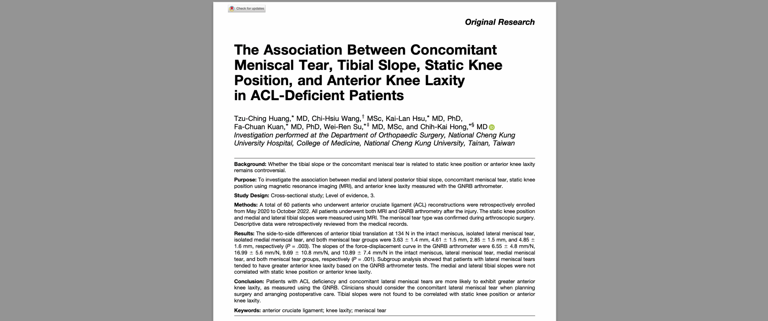
The diagnosis of ACL injuries traditionally relies on physical examination and imaging techniques such as magnetic resonance imaging (MRI). However, these methods are not without limitations. While MRI is widely used for assessing soft tissue injuries, studies have shown its limitations in detecting partial ACL tears . In contrast, robotic arthrometers like the DYNEELAX® and GNRB® provide an objective measurement of knee laxity, which is crucial for identifying ACL ruptures. These devices enable clinicians to conduct dynamic tests that assess both anterior tibial translation and rotational stability, offering a more comprehensive evaluation of knee joint mechanics.
II. The GNRB: A Breakthrough in ACL Injury Diagnosis
The GNRB® arthrometer is specifically designed to measure anterior tibial translation (ATT), a key indicator of ACL integrity. Unlike manual tests such as the Lachman or pivot shift, which can be subject to variability, the GNRB provides quantifiable and reproducible results that are highly accurate in detecting both complete and partial ACL tears.
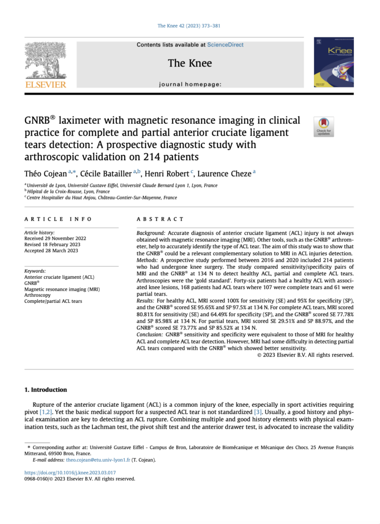
A prospective study by Théo Cojean in 2023 demonstrated that the GNRB®‘s sensitivity and specificity for partial ACL tears were significantly superior to MRI. The GNRB® achieves this by applying graduated forces to the tibia while the knee is in 20° of flexion.
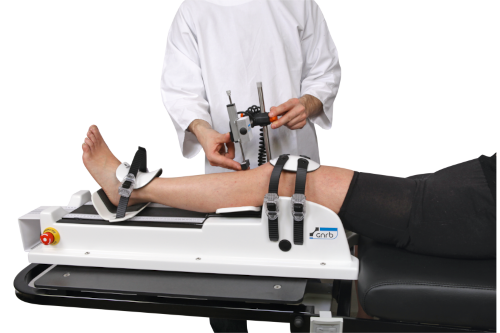
In this study, 214 patients undergoing knee arthroscopy were evaluated with both GNRB® and MRI. The GNRB® outperformed MRI in detecting partial tears, with sensitivity of 73.77% compared to MRI’s 29.51% . This is a critical finding, as partial ACL tears can often go undetected, leading to inadequate treatment and a higher risk of future knee instability or re-injury.
III. Dyneelax: Expanding the Diagnostic Horizon with Rotational Analysis
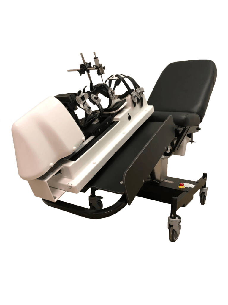
While the GNRB® focuses on anterior translation, the DYNEELAX® arthrometer goes a step further by measuring both anterior tibial translation and rotational instability. This dual measurement capability allows for a more thorough assessment of knee stability, which is particularly important for surgical planning and post-operative rehabilitation.
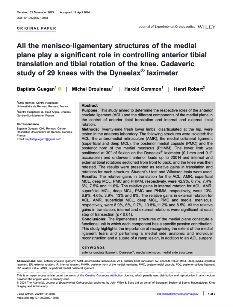
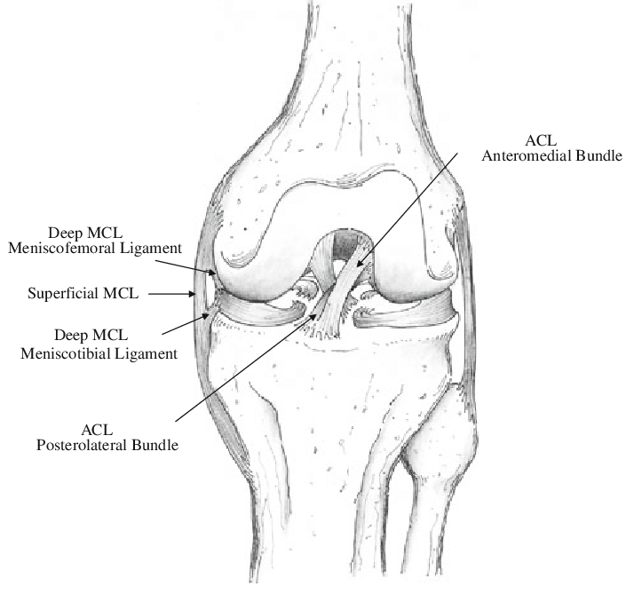
According to a cadaveric study by Guegan et al. (2024), the medial plane structures of the knee, including the medial collateral ligament (MCL) and the posterior medial capsule, play a significant role in controlling both anterior tibial translation and tibial rotation. The DYNEELAX® was instrumental in quantifying the contribution of these structures to knee stability, with results showing that the ACL and surrounding ligaments work synergistically to resist both translational and rotational forces. This data is crucial for surgeons when determining whether additional procedures such as Lateral Extra-Articular Tenodesis (LET) are necessary to address rotational instabilities during ACL reconstruction for example.
IV. Enhancing Surgical Planning and Rehabilitation
The rotational component measured by DYNEELAX® is not only essential for diagnosis but also for surgical planning. For instance, by providing precise data on tibial rotation, Dyneelax helps surgeons identify cases where rotational instability is present, which may require additional procedures like LET. This ensures that the surgical repair addresses all aspects of knee instability, leading to better outcomes and lower re-injury rates.
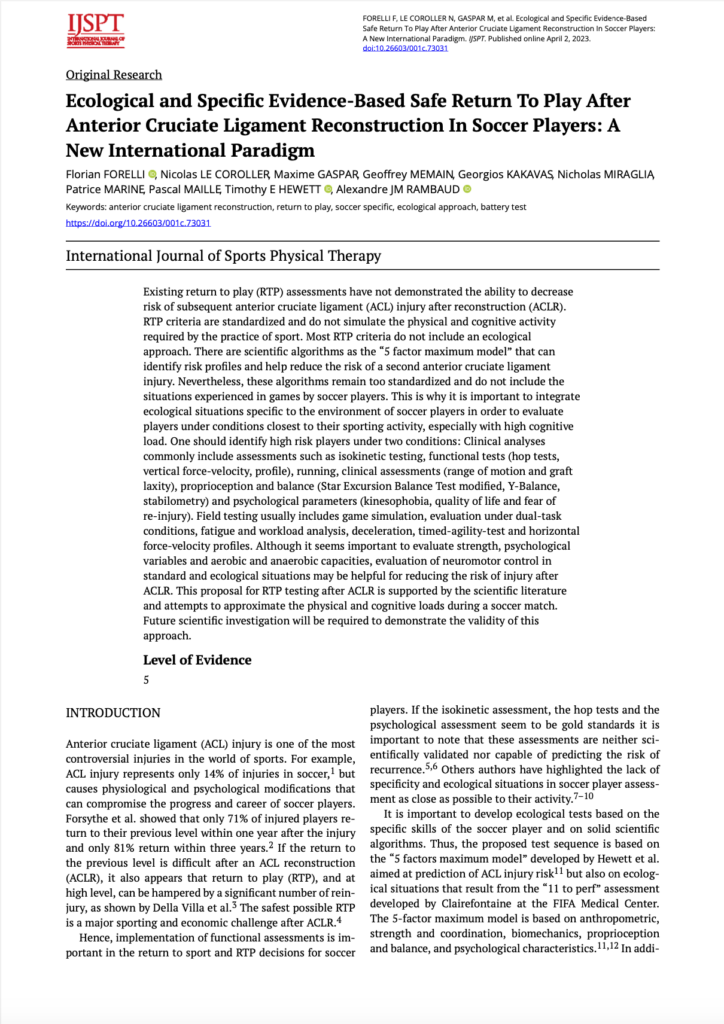
Additionally, DYNEELAX® plays a key role in post-operative monitoring. A study by Forelli et al. (2023) demonstrated the utility of Dyneelax in guiding rehabilitation programs by closely monitoring the stability of the ACL graft post-surgery . This allows for personalized rehabilitation protocols, enabling clinicians to adjust the intensity and type of exercises based on the patient’s recovery progress. By assessing knee stability as early as six weeks post-operation with low-force measurements, Dyneelax provides invaluable data to safely determine when an athlete can return to sport
V. Why Invest in Robotic Arthrometers?
5.1 Financial Return on Investment (ROI)
One of the most compelling reasons for investing in robotic arthrometers is the short return on investment (ROI). Hospitals and clinics that treat a significant number of ACL injuries can quickly recoup the cost of these devices. Based on recent studies, the cost of an MRI in the United States averages around $1,350, while a DYNEELAX® test can be priced at approximately $1,000. Given the higher diagnostic accuracy of DYNEELAX®, patients may opt for this test as a first instance, especially when only ACL injuries are suspected. This could potentially reduce the number of unnecessary MRI scans, freeing up MRI resources for other pathologies .

For example, a hospital seeing 10 ACL patients per month could generate $10,000 per month in revenue from DYNEELAX® testing. With a purchase price signifcantly lower than an MRI machine, the Dyneelax device would pay for itself in less than 2 years, providing a strong financial incentive for healthcare providers . Furthermore, for clinics treating higher volumes of ACL injuries, the ROI could be achieved even more quickly.
5.2 Scientific and Clinical Advantages
In addition to financial benefits, the scientific advantages of GNRB® and DYNEELAX® are undeniable. Both devices provide dynamic, quantitative data that complements traditional static imaging techniques such as MRI. By offering greater accuracy in detecting partial tears, these devices help prevent misdiagnosis and ensure that patients receive the most appropriate treatment from the outset
5.3 Improved Healthcare Efficiency
Finally, the use of robotic arthrometers like DYNEELAX® and GNRB® can lead to more efficient healthcare systems. In countries like France, where MRI wait times can exceed 30 days, using robotic arthrometers as a first-line diagnostic tool for ACL injuries can reduce MRI demand and shorten patient wait times. This improves overall patient care by enabling faster diagnoses and treatment decisions, reducing the burden on healthcare providers
Conclusion: The Future of ACL Injury Diagnosis

The DYNEELAX® and GNRB® robotic arthrometers represent a significant advancement in the diagnosis, treatment planning, and rehabilitation of ACL injuries. By providing objective, reproducible data on both anterior tibial translation and rotational stability, these devices enhance the accuracy of ACL injury diagnosis, particularly for partial tears that may be missed by traditional imaging techniques.
With their proven ability to assist in surgical planning and personalized rehabilitation, as well as their potential to deliver a short ROI, DYNEELAX® and GNRB® are invaluable tools for any clinic or hospital involved in treating knee injuries. Investing in these robotic arthrometers not only improves patient outcomes but also enhances the efficiency of healthcare delivery, making them a wise investment for the future of orthopaedic care.
Medical References (Link in DOI)
- Cojean, T. (2023). MRI vs. GNRB in ACL Injuries Detection: A Comparative Study. Journal of Orthopedic Research and Therapy (DOI: 10.1016/j.knee.2023.03.017).
- Forelli (2023). After Surgery Rehab Post-Op. International Journal of Sports Physical Therapy. Submitted: October 10, 2022. Accepted: January 15, 2023. DOI: 10.26603/001c.73031.
- Guegan, B., Drouineau, M., Common, H., & Robert, H. (2024). All the menisco-ligamentary structures of the medial plane play a significant role in controlling anterior tibial translation and tibial rotation of the knee: Cadaveric study of 29 knees with the Dyneelax® laximeter. Journal of Experimental Orthopaedics, 11, e12038. DOI: 10.1002/jeo2.12038

