A thorough knee examination is essential for diagnosing ligament injuries, meniscal tears, and joint instability. Orthopaedic specialists rely on clinical tests and advanced diagnostic tools to evaluate knee function accurately. A structured approach to knee examination includes assessing patient history, performing a physical examination, and utilizing advanced diagnostic methods. This article explores these key aspects, emphasizing the importance of manual and instrumented assessments before discussing modern advancements in knee laxity evaluation.
I. Why a Thorough Knee Examination Matters
A precise knee examination is fundamental for identifying conditions such as ligament tears, meniscus injuries, osteoarthritis, and patellar instability. Without a structured assessment, minor injuries may go unnoticed, leading to chronic instability and degenerative changes over time.
A knee examination serves several purposes:
Identifying the underlying cause of knee pain or instability.
Determining the severity of injury to guide treatment planning.
Assessing functional limitations in athletes and active individuals.
Monitoring post-operative recovery and rehabilitation progress.
By integrating a standardized knee examination into daily clinical practice, healthcare providers can ensure accurate diagnoses and optimal patient outcomes.
II. Clinical Assessment of the Knee
The clinical examination of the knee involves a systematic approach to evaluating pain, range of motion, stability, and ligament integrity. The main components include:
1. Patient History
A detailed history provides crucial insights into the mechanism of injury and symptoms. Important questions include:
When did the pain or instability start?
Was there a traumatic event or sudden twisting motion?
Are there episodes of the knee “giving way”?
Any previous surgeries or underlying conditions affecting knee stability?
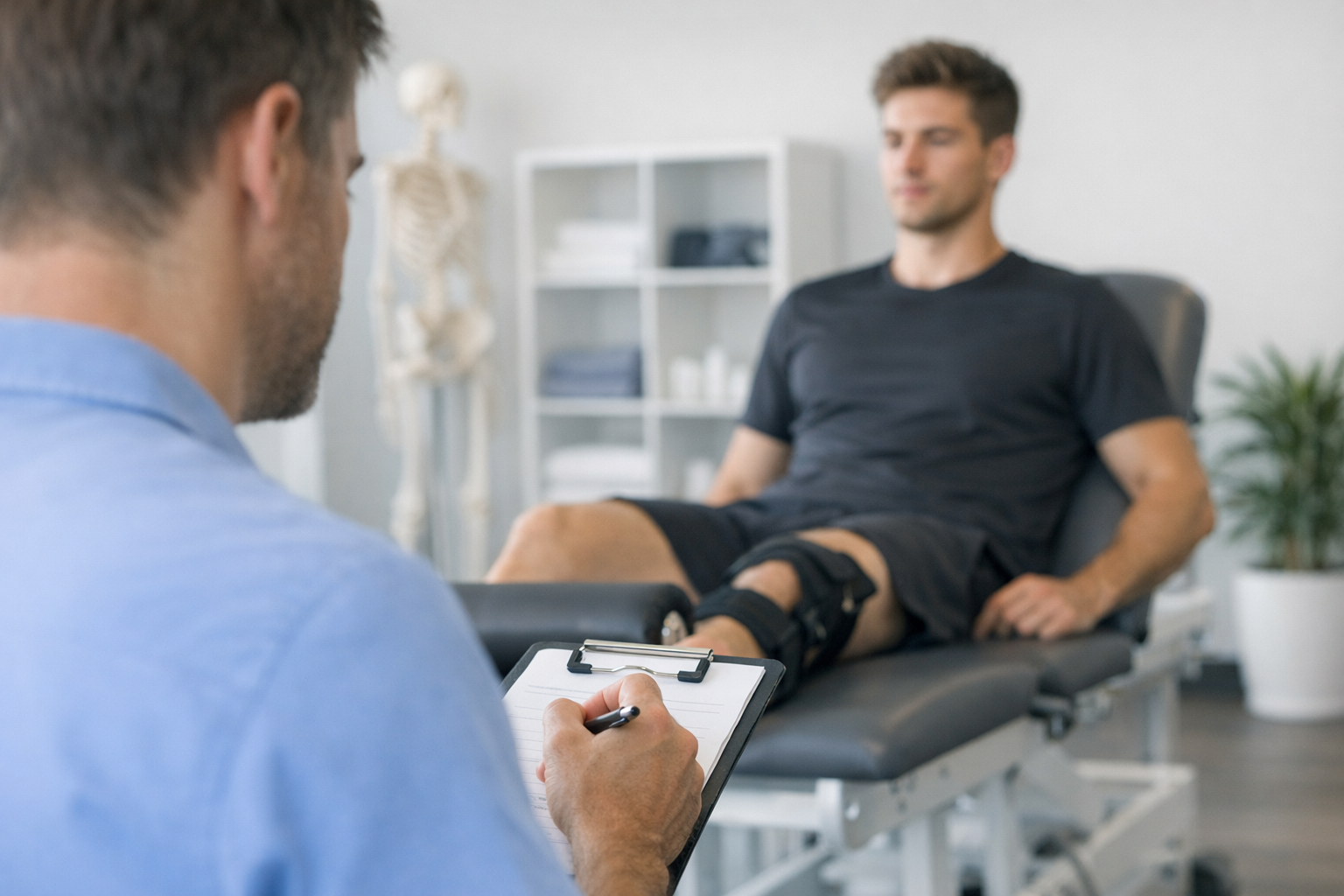
2. Inspection and Palpation
Visual inspection is essential for identifying swelling, deformities, and muscle atrophy. Palpation helps locate tenderness over key structures such as the medial and lateral joint lines, patella, and tibial tuberosity.
3. Range of Motion (ROM) Testing
Active and passive ROM tests assess the knee’s flexibility and any mechanical blockages. A normal knee should extend fully (0°) and flex up to approximately 135°.
4. Manual Knee Stability Tests
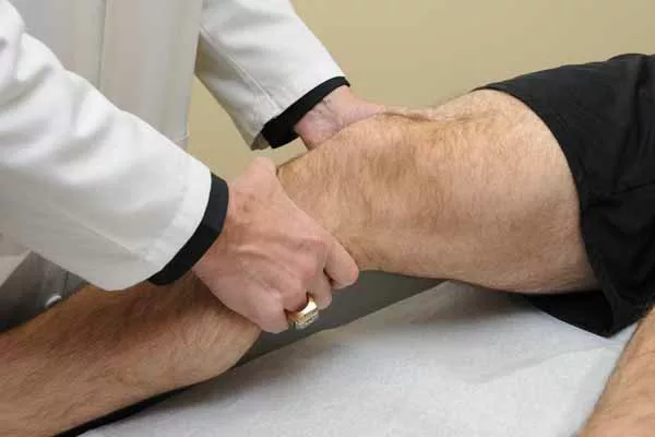
To assess ligament integrity, orthopaedic specialists perform several key tests:
Anterior Drawer Test: Evaluates anterior cruciate ligament (ACL) laxity.
Lachman Test: More sensitive than the anterior drawer test for ACL injuries.
Pivot Shift Test: Detects rotational instability in ACL-deficient knees.
Valgus and Varus Stress Tests: Assess medial and lateral collateral ligament stability.
Posterior Drawer Test: Checks posterior cruciate ligament (PCL) integrity.
While these tests provide useful clinical insights, manual examinations can be subjective and dependent on examiner experience. This limitation has led to the development of robotic knee arthrometers for objective knee laxity assessment.
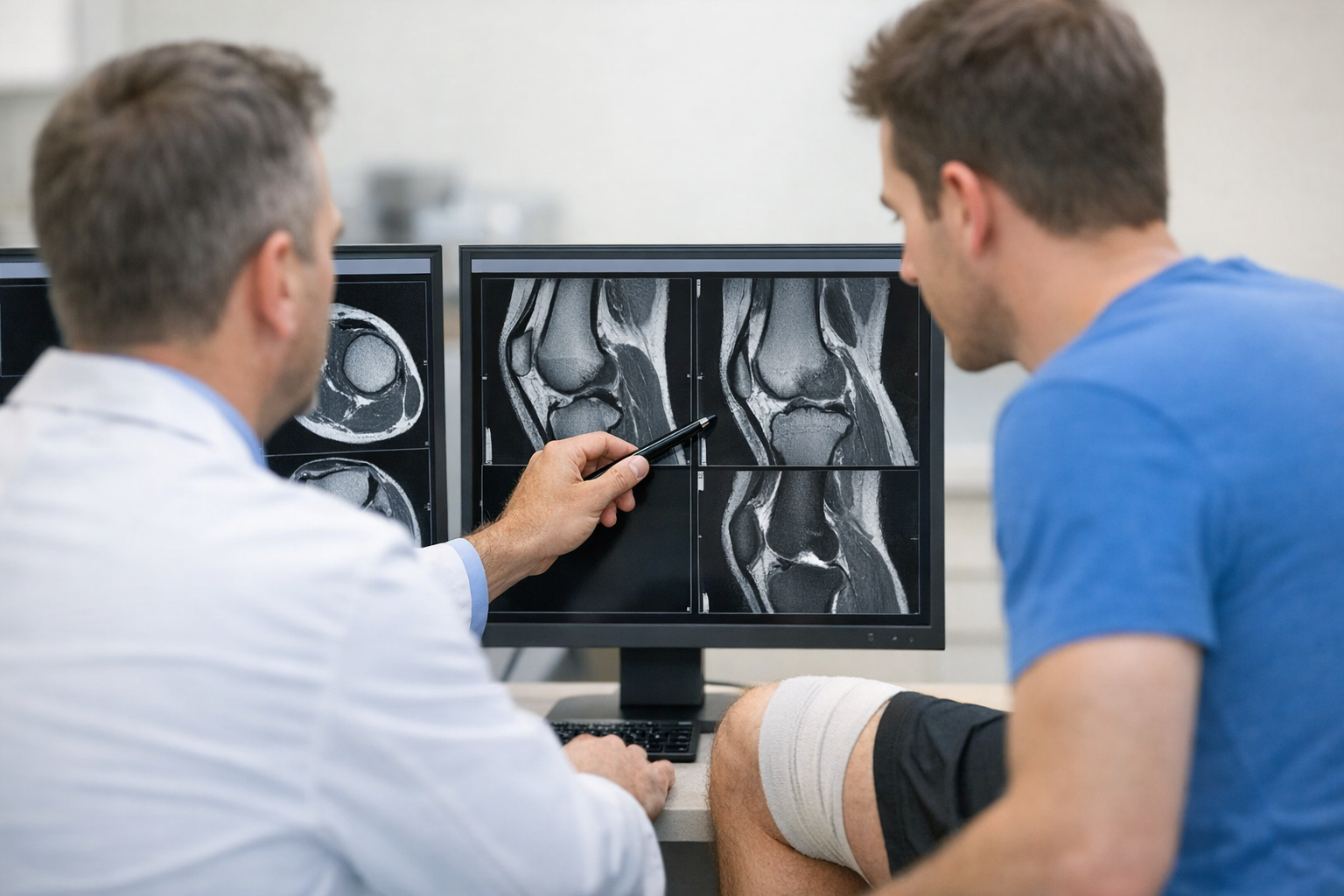
III. The Role of Robotic Arthrometers in Knee Laxity Evaluation
Recent research has reinforced the importance of robotic arthrometry in knee laxity evaluation. Studies have demonstrated that robotic devices such as DYNEELAX® and GNRB® provide superior accuracy compared to traditional manual tests, making them invaluable in ACL and multi-ligament injury assessments.
A 2023 study by Cojean et al. evaluated the GNRB laximeter’s effectiveness in detecting complete and partial ACL tears compared to MRI. The study, which included 214 patients, found that GNRB had higher sensitivity for partial ACL tears and comparable accuracy to MRI for complete ACL ruptures, reinforcing its role as a complementary diagnostic tool (Cojean et al., 2023).
Another study by Pouderoux et al. (2019) focused on the evolution of joint laxity and graft compliance following ACL reconstruction. Using the GNRB arthrometer, they tracked changes in knee laxity over time, demonstrating that robotic arthrometry is highly useful for monitoring post-operative recovery and determining the optimal timing for return to sports (Pouderoux et al., 2019).
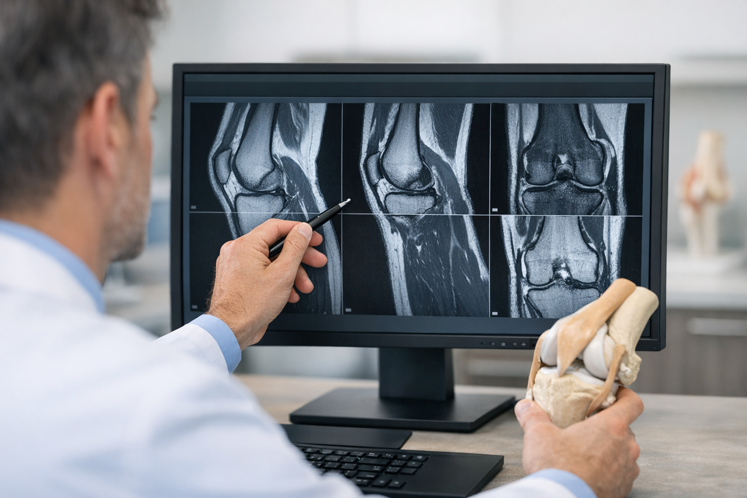
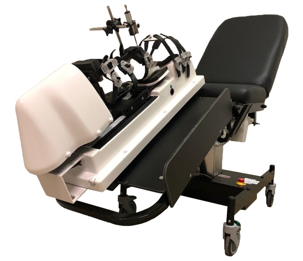
The Dyneelax laximeter has further revolutionized multi-parameter knee laxity evaluation by measuring both anterior tibial translation and rotational stability. A 2024 cadaveric study by Guegan et al. examined the role of medial plane structures in controlling tibial translation and rotation using the Dyneelax laximeter. The study highlighted the importance of assessing medial ligament contributions to knee stability and emphasized Dyneelax’s role in identifying complex ligamentous injuries (Guegan et al., 2024).
Knee laxity is a crucial parameter in diagnosing ligamentous injuries. Traditional manual tests can be inconsistent due to examiner variability. The introduction of robotic arthrometers, such as DYNEELAX® and GNRB®, has revolutionized knee laxity measurement by providing quantifiable, reproducible, and highly accurate assessments.
1. GNRB: Precise ACL Laxity Measurement
The GNRB® robotic arthrometer is specifically designed to evaluate anterior tibial translation, the primary indicator of ACL laxity. Unlike manual tests, GNRB® applies controlled, progressive anterior forces while tracking tibial displacement in real-time. Key advantages include:
-
Objective measurement of ACL laxity without examiner bias.
-
Comparison with the contralateral knee for precise diagnosis.
-
Quantitative data to track post-operative recovery.
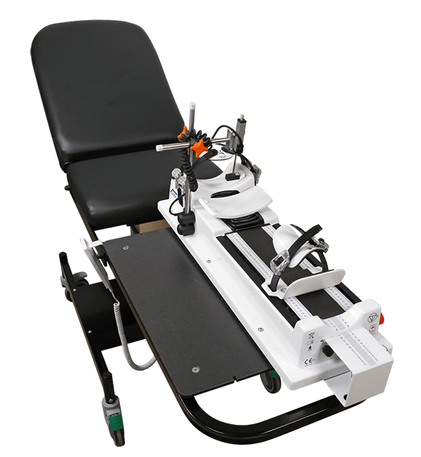
2. Dyneelax: A Multi-Parameter Knee Laxity Analyzer
IV. Benefits of Robotic Knee Examination
The adoption of robotic knee arthrometry enhances knee examinations by:
Providing numerical data for ligament evaluation, eliminating subjectivity.
Detecting early-stage instability, which may be missed in manual tests.
Supporting surgical planning by differentiating between isolated ACL injuries and combined ligament damage.
Monitoring rehabilitation progress in athletes and post-surgical patients.
V. Integrating Robotic Arthrometry in Clinical Practice
Conclusion
A comprehensive knee examination requires both traditional clinical methods and advanced robotic technology. While manual tests remain a fundamental part of orthopaedic assessment, robotic arthrometers like DYNEELAX® and GNRB® provide an unparalleled level of precision in knee laxity measurement. By integrating these tools, clinicians can ensure more accurate diagnoses and improved patient care in knee ligament injuries.
Medical References
Cojean T, Batailler C, Robert H, Cheze L. GNRB laximeter with magnetic resonance imaging in clinical practice for complete and partial anterior cruciate ligament tears detection: A prospective diagnostic study with arthroscopic validation on 214 patients. The Knee. 2023;42:373–381. https://doi.org/10.1016/j.knee.2023.03.017.
Pouderoux T, Muller B, Robert H. Joint laxity and graft compliance increase during the first year following ACL reconstruction with short hamstring tendon grafts. Knee Surgery, Sports Traumatology, Arthroscopy. 2019;27(8):2712–2719. https://doi.org/10.1007/s00167-019-05711-z.
Guegan B, Drouineau M, Common H, Robert H. All the menisco-ligamentary structures of the medial plane play a significant role in controlling anterior tibial translation and tibial rotation of the knee. Cadaveric study of 29 knees with the Dyneelax® laximeter. J Exp Orthop. 2024;11:e12038. https://doi.org/10.1002/jeo2.12038.