Understanding the specific roles of the various ligamentous structures within the knee is essential for improving clinical outcomes in patients with knee injuries. A recent study titled “All the menisco‐ligamentary structures of the medial plane play a significant role in controlling anterior tibial translation and tibial rotation of the knee” aimed to shed light on this topic. This study, conducted using the Dyneelax® laximeter/arthrometer, provides critical insights into the biomechanics of the knee, particularly focusing on the anterior cruciate ligament (ACL) and the medial structures of the knee.
The purpose of the study was to determine the respective contributions of the ACL and the medial plane components in controlling anterior tibial translation (ATT) and both internal and external tibial rotation. By utilizing a precise and validated device like the Dyneelax® laximeter/arthrometer, which offers high accuracy in measuring knee movements, the study was able to provide detailed data on how each structure within the knee contributes to its overall stability.
Such research is crucial as it informs surgical strategies and rehabilitation protocols, particularly in complex knee injuries where multiple structures may be involved. The findings underscore the importance of comprehensive diagnostic tools like the Dyneelax® laximeter/arthrometer and GNRB laximeter/arthrometer, which help clinicians assess and manage knee stability more effectively. This article will delve into the methodologies, findings, and clinical implications of this significant study, highlighting its contribution to the field of orthopedic surgery and sports medicine.
I. Study Overview
1.1 Objective of the Study
The primary aim of the study was to determine the specific roles of the anterior cruciate ligament (ACL) and the various components of the medial plane in controlling knee movements, particularly anterior tibial translation (ATT) and both internal and external tibial rotation. Understanding these roles is essential for enhancing clinical approaches to diagnosing and treating knee injuries, especially those involving multiple ligamentous structures.
1.2 Methods Overview
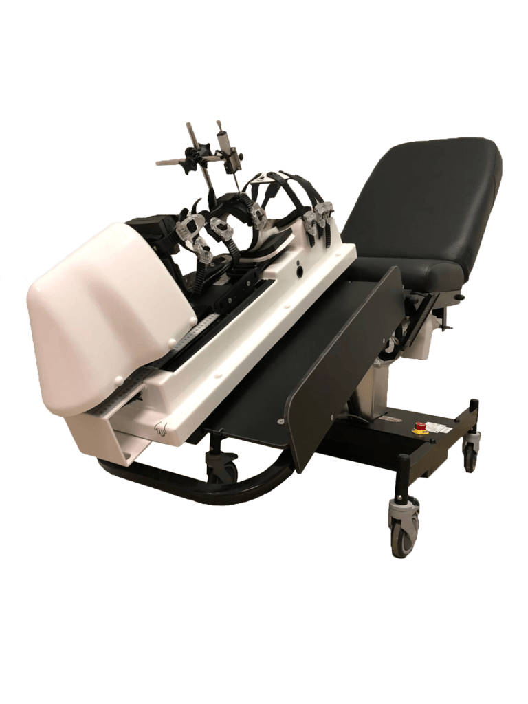
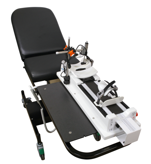
The study was conducted using 29 fresh cadaveric lower limbs, which were disarticulated at the hip and tested in a controlled laboratory environment. Each specimen was meticulously prepared to ensure the integrity of the ligamentous structures before testing. The Dyneelax® laximeter/arthrometer, a highly accurate and validated device, was employed to measure the knee movements. This laximeter/arthrometer allowed for precise measurement of ATT and rotational movements under controlled loads, providing detailed data on the contributions of each knee structure.
Sequential transections were performed on the ACL, anteromedial retinaculum (AMR), superficial medial collateral ligament (sMCL), deep medial collateral ligament (dMCL), posterior medial capsule (PMC), and the posterior horn of the medial meniscus (PHMM). The Dyneelax® laximeter/arthrometer recorded the resulting changes in knee laxity after each transection. Statistical analyses, including Student’s t-tests and Wilcoxon tests, were conducted to determine the significance of the findings.
This methodology enabled a comprehensive evaluation of the passive contributions of each structure to knee stability, offering valuable insights for clinical practice and surgical planning.
II. Materials and Methods
2.1 Study Design
The study was designed to investigate the roles of the anterior cruciate ligament (ACL) and the medial plane components in controlling knee movements. A total of 29 fresh cadaveric lower limbs were utilized, with 17 specimens from females and 12 from males, aged between 41 and 92 years, averaging 81 years. These limbs were disarticulated at the hip and preserved at -20°C until the time of the study. Prior to testing, each specimen was thawed at room temperature and mobilized to ensure flexibility and stability.
2.2 Dissection Technique
An experienced orthopedic surgeon performed all dissections. The medial side of each knee was exposed through a long medial arciform incision, taking care to preserve the ligaments and joint capsule. The dissection process involved identifying and isolating the ACL, anteromedial retinaculum (AMR), superficial medial collateral ligament (sMCL), deep medial collateral ligament (dMCL), posterior medial capsule (PMC), and the posterior horn of the medial meniscus (PHMM). Each structure was meticulously prepared, with No. 2 Vicryl sutures placed around them to aid in transection. The whole ACL was dissected at the midportion through an anteromedial vertical arthrotomy.
2.3 Testing Protocol
The testing protocol involved the use of the Dyneelax® laximeter/arthrometer to measure anterior tibial translation (ATT) and rotational movements. Each knee was positioned at 30° of flexion on the Dyneelax® laximeter/arthrometer, with the femur secured to prevent rotation and the foot and ankle attached to a stationary block. The initial measurements of knee movements were recorded with all structures intact.
Sequential transections were then performed in the following order: ACL, AMR, sMCL, dMCL, PMC, and PHMM. After each transection, the knee was retested using the Dyneelax® laximeter/arthrometer to measure changes in ATT and rotational movements. The results were recorded as relative gains in translation and rotation for each structure, with statistical analyses conducted to determine the significance of the changes observed.
This detailed methodology ensured precise and reproducible measurements of knee stability, providing valuable insights into the contributions of each ligamentous structure.
III. Materials and Methods
3.1 Key Findings
The study yielded significant insights into the roles of various knee structures in controlling anterior tibial translation (ATT), internal rotation (IR), and external rotation (ER). The anterior cruciate ligament (ACL) was found to be the primary restraint for ATT, showing a relative gain of 42.9% in translation control after its transection. For IR, the ACL exhibited a relative gain of 13%, while for ER, it showed a gain of 8.9%.
The anteromedial retinaculum (AMR) demonstrated a 6.7% relative gain in ATT control, a 6.9% gain in IR control, and a 6% gain in ER control after transection. The superficial medial collateral ligament (sMCL) showed a 7.4% relative gain in ATT, 4.6% in IR, and 9.7% in ER. The deep medial collateral ligament (dMCL) exhibited gains of 6% in ATT, 3.9% in IR, and 13.8% in ER. The posterior medial capsule (PMC) had a significant role in controlling IR with a relative gain of 13%, and also showed gains of 7.5% in ATT and 11.2% in ER. Finally, the posterior horn of the medial meniscus (PHMM) contributed 11.6% in ATT control, 8% in IR, and 8.5% in ER.
3.2 Statistical Analysis
The statistical analysis confirmed the significance of the observed changes in knee stability after the transection of each structure. All relative gains in translation, internal, and external rotations were found to be significant at each step of transection (p < 0.01).
- ACL: The ACL’s transection resulted in a significant increase in ATT (2.9 mm), IR (1.4°), and ER (1°).
- AMR: The AMR’s removal showed significant increases in ATT (0.5 mm), IR (0.8°), and ER (0.7°).
- sMCL: After transecting the sMCL, significant increases were noted in ATT (0.5 mm), IR (0.5°), and ER (1.4°).
- dMCL: The dMCL transection led to significant increases in ATT (0.5 mm), IR (0.5°), and ER (1.8°).
- PMC: Removing the PMC resulted in significant increases in ATT (0.5 mm), IR (1.5°), and ER (1.3°).
- PHMM: The PHMM’s transection showed significant gains in ATT (0.9 mm), IR (0.7°), and ER (0.8°).
Overall, the study highlighted the critical roles that each of these structures plays in maintaining knee stability. The findings emphasize the importance of considering all these components when diagnosing and treating knee injuries, particularly in the context of ACL and medial ligament reconstructions. This comprehensive analysis using the Dyneelax® laximeter/arthrometer provides a clearer understanding of knee biomechanics and offers valuable data for clinical applications.
IV. Discussion
4.1 Interpretation of Results
The study provides a detailed analysis of the roles played by the anterior cruciate ligament (ACL) and various medial knee structures in controlling anterior tibial translation (ATT), internal rotation (IR), and external rotation (ER). The ACL was confirmed as the primary restraint for ATT, with a significant relative gain of 42.9% in translation control after transection. This finding aligns with previous studies that have consistently shown the ACL’s crucial role in preventing anterior displacement of the tibia.
The anteromedial retinaculum (AMR), though less studied, demonstrated notable contributions to IR (6.9%) and ER (6%), supporting the hypothesis that it plays a significant role in rotational stability. The superficial medial collateral ligament (sMCL) was primarily involved in controlling ER (9.7%), which corroborates earlier research indicating its importance in valgus and rotational stability.
The deep medial collateral ligament (dMCL) exhibited the highest relative gain in ER control (13.8%), highlighting its role as a primary restraint in this motion, similar to findings in biomechanical studies that emphasize the dMCL’s importance in preventing excessive external rotation. The posterior medial capsule (PMC) showed major contributions to IR (13%), consistent with its anatomical positioning and function in providing internal rotational stability.
Finally, the posterior horn of the medial meniscus (PHMM) demonstrated secondary but significant roles in ATT (11.6%) and rotational control, supporting the concept of the meniscus acting as a stabilizing structure in the knee.
4.2 Clinical Implications
The findings of this study have significant clinical implications for the diagnosis and treatment of knee injuries. Understanding the distinct roles of these medial knee structures can aid in the accurate diagnosis of complex ligament injuries, particularly in cases involving combined ACL and medial ligament tears. The use of the Dyneelax® laximeter/arthrometer in this study highlights the importance of precise diagnostic tools in evaluating knee stability and planning surgical interventions.
For clinicians, these insights emphasize the need for comprehensive assessments of all medial structures when addressing knee instability. In ACL reconstruction surgeries, attention must be given to associated injuries in the medial structures to prevent residual instability and improve surgical outcomes. For instance, recognizing the significant role of the dMCL in ER control suggests that reconstructing this ligament alongside the ACL could be crucial in patients with anteromedial rotatory instability (AMRI).
Moreover, the study underscores the importance of repairing ramp lesions of the PHMM to restore knee stability fully. This comprehensive approach to diagnosing and treating knee injuries ensures that all contributing factors to instability are addressed, leading to better patient outcomes and reduced risk of re-injury.
In summary, the study’s detailed analysis using the Dyneelax® laximeter/arthrometer provides valuable data that enhance our understanding of knee biomechanics and inform clinical practices in orthopedic surgery and sports medicine.
Conclusion
The study using the Dyneelax® laximeter/arthrometer revealed critical insights into the roles of the anterior cruciate ligament (ACL) and medial knee structures in controlling anterior tibial translation (ATT), internal rotation (IR), and external rotation (ER). The ACL was identified as the primary restraint for ATT, while the deep medial collateral ligament (dMCL) was crucial for ER control, and the posterior medial capsule (PMC) for IR. These findings highlight the importance of a comprehensive approach in diagnosing and treating knee injuries, emphasizing the need to address all involved structures to restore stability fully.
Future research should focus on dynamic testing of knee structures under various flexion angles to simulate real-life movements more accurately. Additionally, exploring the roles of other ligaments and soft tissue structures in knee stability could further enhance surgical techniques and rehabilitation protocols. Improved diagnostic tools and individualized reconstruction strategies could lead to better patient outcomes and reduced rates of re-injury.
Impact on Arthrometers
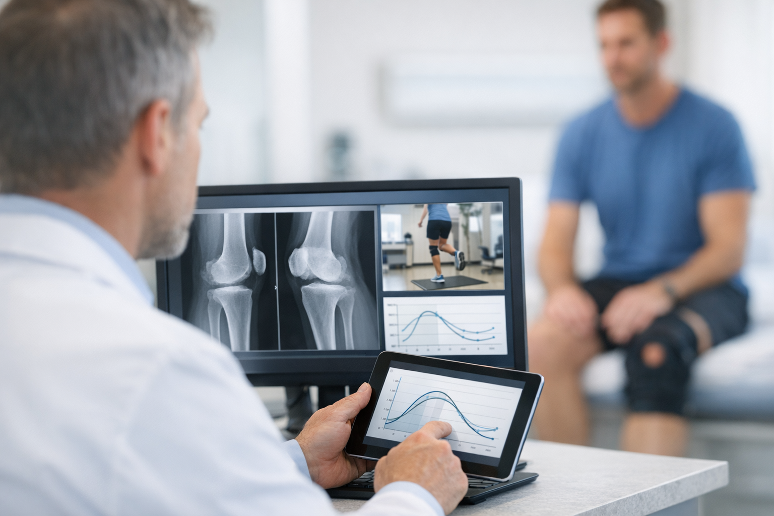
The findings of this study strongly support the use of GNRB and Dyneelax® laximeters/arthrometers in clinical settings for diagnosing and managing knee instability. The precise measurements provided by the Dyneelax® laximeter/arthrometer were crucial in identifying the specific contributions of various knee structures to anterior tibial translation (ATT), internal rotation (IR), and external rotation (ER).
Similarly, the GNRB laximeter/arthrometer is renowned for its accuracy in assessing ACL integrity and overall knee stability, making it an invaluable tool for orthopedic surgeons and sports medicine specialists.
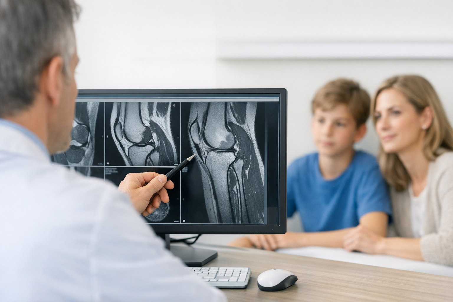
The primary advantages of using GNRB and Dyneelax® laximeters/arthrometers include their high precision, reproducibility, and ability to provide objective data on knee laxity. These devices offer measurements with up to 0.1 mm and 0.1° accuracy, allowing clinicians to detect subtle changes in knee stability that may not be apparent through manual testing alone. Notably, these laximeters/arthrometers can detect partial ruptures more effectively than traditional MRI scans (Cojean Study – 2023), which often miss such nuanced injuries. This enhanced detection capability is crucial for early and accurate diagnosis, leading to more effective treatment plans.
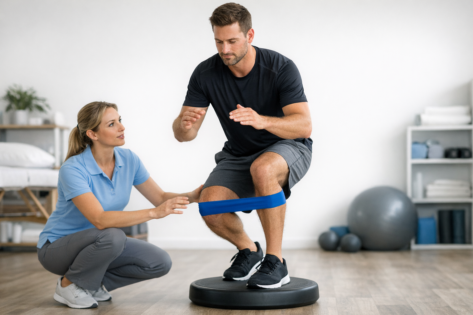
Additionally, the non-invasive nature of these laximeters/arthrometers ensures patient comfort and safety during assessment. One significant benefit is the ability to monitor the graft (Forelli study – 2023) post-operatively in patients who have undergone ACL reconstruction. By providing precise measurements of knee stability over time, clinicians can track the progress of graft healing and ensure that the knee is recovering as expected, allowing for timely interventions if any issues arise.
The ability to quantify knee movements accurately aids in the early detection of instability and the evaluation of treatment efficacy, ultimately leading to better patient outcomes. As demonstrated by this study, integrating these advanced diagnostic tools into clinical practice enhances our understanding of knee biomechanics and improves the management of complex knee injuries.
Medical References
- Guegan, B., Drouineau, M., Common, H., & Robert, H. (2024). All the menisco-ligamentary structures of the medial plane play a significant role in controlling anterior tibial translation and tibial rotation of the knee: Cadaveric study of 29 knees with the Dyneelax® laximeter. Journal of Experimental Orthopaedics, 11, e12038. DOI: 10.1002/jeo2.12038
- Cojean, T. (2023). MRI vs. GNRB in ACL Injuries Detection: A Comparative Study. Journal of Orthopedic Research and Therapy (DOI: 10.1016/j.knee.2023.03.017).
- Forelli F, Le Coroller N, Gaspar M, et al. (2023). Ecological and Specific Evidence-Based Safe Return To Play After Anterior Cruciate Ligament Reconstruction In Soccer Players: A New International Paradigm. IJSPT. Published online April 2, 2023. DOI: 10.26603/001c.73031