Study Title: Is the Lind or LaPrade Technique Able to Control Anterior and Rotational Laxity in Medial-Side Knee Injuries?
Authors:
Michel Drouineau, MD, Baptiste Guegan, MD, Harold Common, MD, Theo Cojean, PhD, and Henri Robert, MD
Journal: The Orthopaedic Journal of Sports Medicine (OJSM)
Publication Date: 2025
DOI: doi.org/10.1177/23259671251389124
Institution: Centre Hospitalier Universitaire de Rennes, Rennes, France
Medial-side knee injuries are often discussed as “MCL tears,” but the reality is more complex. When the medial collateral ligament (MCL) is compromised together with posteromedial structures like the posterior oblique ligament (POL), the knee can develop a combination of valgus, anterior, and rotational laxity. That mix is clinically important because it can contribute to persistent instability, overload secondary stabilizers, and potentially jeopardize outcomes in combined ligament scenarios.
A late-2025 paper by Drouineau et al. (Orthopaedic Journal of Sports Medicine) took a very practical question and tested it in a controlled lab setting: between two classic medial-side reconstructions, the Lind and the LaPrade techniques, which one better controls anterior tibial translation and rotational laxity after a severe medial injury?
Key points in 30 seconds
The study simulated a grade 3 medial-side injury (MCL + POL deficiency) on 18 cadaveric knees.
Both reconstructions improved stability compared with the injured state.
Lind showed less residual anterior translation and less residual external rotation laxity than LaPrade (statistically significant in absolute values).
Neither technique fully restored native translation and rotation at “time zero,” suggesting room for technical refinement.
Why this question matters
In daily practice, medial laxity is not only a “side-to-side opening” problem. The medial structures also contribute to controlling tibial rotation and anterior translation, especially in combined patterns such as anteromedial rotatory instability (AMRI). If a reconstruction restores valgus stability but leaves subtle rotational laxity, patients may still report giving way, and surgeons may continue to observe borderline pivoting or persistent subjective instability.
That is why comparing “classic” reconstructions is more than a technical curiosity: it helps clarify what each construct actually controls biomechanically.
Study design: controlled cadaveric testing
This was a controlled laboratory study using 18 fresh-frozen cadaveric lower limbs (mean donor age around the mid-70s). Each knee was tested in a consistent setup at 30° of flexion.
The authors created a stepwise model to observe how laxity evolves as structures are cut and then reconstructed:
Intact knee
MCL transection (superficial + deep layers in their dissection approach)
MCL + POL transection (to simulate a severe medial-side injury)
Reconstruction with either:
Lind technique (9 knees), or
LaPrade technique (9 knees)
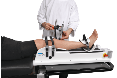
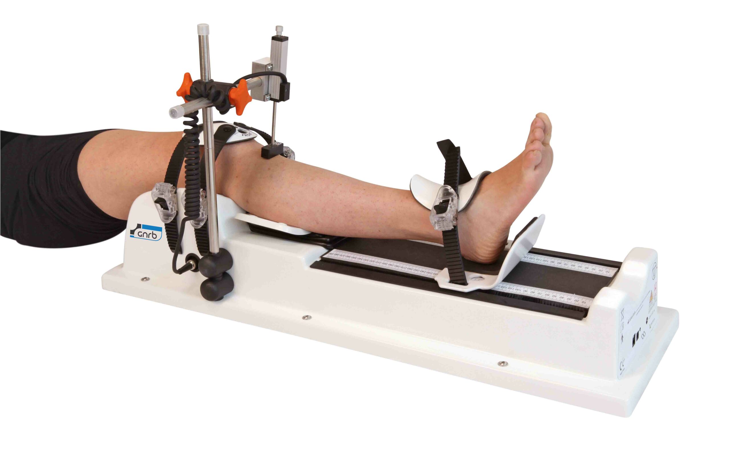
To quantify stability, they used a combined translation/rotation testing device (a knee laximeter) able to apply:
An anterior force up to the testing level (reported at 200 N for analysis), and
Internal and external rotation torque (reported at 5 N·m for analysis)
Measurements were repeated multiple times to reduce noise, and the study reported both:
Absolute residual laxity (how far from intact, in mm or degrees), and
Relative residual laxity (how much of the injury-induced laxity is “corrected,” in %).
A quick refresher: what are the Lind and LaPrade techniques?
Both procedures aim to address medial and posteromedial deficiency by reconstructing key restraints, but they do it differently.
Lind technique (2009 concept)
In simplified terms, this approach reconstructs the medial side using a single main graft path to re-create control of both MCL and POL function with fewer tunnels. In the study, a semitendinosus graft was used with fixation designed to create an inverted “V” appearance on the medial side.
LaPrade technique (anatomic medial reconstruction concept)
The LaPrade approach is typically described as a more anatomic reconstruction, rebuilding both the superficial MCL (sMCL) and POL with distinct graft constructs and attachment points (often using semitendinosus for sMCL and gracilis for POL in lab setups).
Because surgeons often choose one technique based on training, preference, or perceived anatomic fidelity, it is valuable to compare them under identical injury conditions and identical testing loads.
What happened after cutting the medial structures?
As expected, transecting the medial restraints increased laxity.
In both groups, the biggest jump occurred when moving from isolated MCL deficiency to combined MCL + POL deficiency, especially for rotational measures. That fits the idea that the posteromedial structures play an important role in controlling rotational behavior when the knee is loaded.
Main results: residual laxity after reconstruction
This is the heart of the paper: after reconstructing the medial side, how close did each technique come to the intact knee?
Anterior tibial translation (ATT) at 200 N
-
Lind: residual laxity about 0.70 mm
-
LaPrade: residual laxity about 1.21 mm
-
Lind was statistically better in absolute ATT control.
Internal rotation (IR) at 5 N·m
-
Lind: residual laxity about 0.92°
-
LaPrade: residual laxity about 0.98°
-
No meaningful difference between techniques here.
External rotation (ER) at 5 N·m
-
Lind: residual laxity about 0.48°
-
LaPrade: residual laxity about 1.21°
-
Lind was statistically better in absolute ER control.
One nuance the authors highlight: while absolute differences reached statistical significance for ATT and ER, differences in relative correction (%) were less striking. In other words, both techniques “fixed most of the problem,” but Lind tended to end closer to intact, especially for external rotation.
How should clinicians interpret these numbers?
A key practical interpretation offered by the authors is that the remaining laxity, although measurable with instrumentation, may be small enough to be difficult to detect clinically in a standard manual exam, particularly at time zero. This is exactly why instrumented laxity measurements can add value: they reveal subtle differences that hands alone might miss.
At the same time, the conclusion is not “Lind wins, full stop.” The more important message is:
-
Both reconstructions improve stability compared with the injured state.
-
Neither fully restores native translation and rotation immediately after surgery (in this lab model).
So the study supports the concept that we still have a “biomechanical gap” in medial-side reconstructions, especially if the goal is to reproduce intact kinematics rather than simply reduce gross instability.
Why might neither technique fully restore native stability?
The discussion points toward several plausible reasons (and these are relevant to everyday surgical reasoning):
The native sMCL is broad and flat, with complex fiber behavior, while many reconstructions use round, cord-like grafts that may not reproduce that function perfectly.
Medial stability is not purely passive. Dynamic contributors (musculotendinous support) are absent in cadaveric models.
Testing was performed at a single flexion angle (30°) and under standardized loads, which is excellent for comparison but cannot represent every functional position or real-life movement pattern.
Study limitations to keep in mind
As with all cadaveric “time-zero” biomechanics, there are constraints:
Older donor tissue may not behave like young athletic tissue.
No healing, no biological remodeling, and no post-op rehab effects are represented.
The model isolates passive structures and does not include muscle activation.
The findings apply to the specific testing setup and fixation choices used in the lab.
These limitations do not invalidate the results—they simply define what the paper can (and cannot) claim.
Take-home message
If you want a one-sentence summary: both Lind and LaPrade medial reconstructions improve sagittal and rotational stability after a severe medial-side injury, but Lind achieved smaller residual anterior translation and external rotation laxity in this controlled cadaveric model.
The bigger takeaway is also forward-looking: even widely used “classic” techniques may still leave a measurable gap versus native stability, which supports ongoing innovation in medial-side reconstruction design, graft geometry, fixation strategy, and perhaps more comprehensive restoration of the medial complex.