Study Title: The Association Between Concomitant Meniscal Tear, Tibial Slope, Static Knee Position, and Anterior Knee Laxity in ACL-Deficient Patients
Authors: Tzu-Ching Huang, Chi-Hsiu Wang, Kai-Lan Hsu, Fa-Chuan Kuan, Wei-Ren Su, Chih-Kai Hong
Journal: Orthopaedic Journal of Sports Medicine (OJSM)
Publication Date: March 2025
DOI: 10.1177/23259671251324186
Institution: Department of Orthopaedic Surgery, National Cheng Kung University Hospital, Taiwan
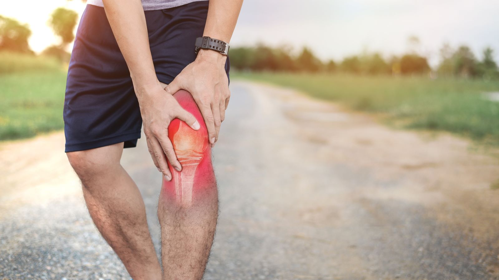
A new peer-reviewed clinical study has brought valuable insights into how ACL injuries are biomechanically influenced by associated meniscal pathology. Leveraging the GNRB® robotic arthrometer and MRI, this study analyzed 60 patients with ACL ruptures and revealed that lateral meniscal tears are strongly associated with increased anterior knee laxity.
The findings further confirm the clinical utility of GNRB in objectively quantifying both anterior translation and stiffness, offering significant advantages over older methods such as the KT-1000 or Telos.
Key Takeaways
Patients with ACL + lateral meniscal tears exhibited significantly greater anterior tibial translation and lower ligamentous stiffness than other groups.
GNRB® provided reliable, reproducible, and precise measurement of knee laxity and stiffness in vivo.
Tibial slope, as measured by MRI, was not associated with either static knee position or anterior knee laxity.
Clinical management of ACL injuries should account for the amplified instability caused by lateral meniscal tears, confirmed via robotic arthrometry.
Study Design & Methodology
Type: Cross-sectional, Level 3 evidence
Period: May 2020 – October 2022
Sample Size: 60 ACL-deficient patients
Tools Used:
MRI (for static knee position and tibial slope)
GNRB® robotic arthrometer (for anterior tibial translation and stiffness analysis)
Patients were grouped into four categories based on arthroscopically confirmed meniscal tear patterns:
No meniscal tear (n = 17)
Lateral meniscal tear only (n = 14)
Medial meniscal tear only (n = 16)
Both medial and lateral tears (n = 13)
Main Results
Increased Laxity in Lateral Meniscal Tear Patients
The side-to-side differences (SSD) in anterior tibial translation under a 134 N load were:
No meniscal tear: 3.63 ± 1.4 mm
Lateral meniscal tear: 4.61 ± 1.5 mm
Medial meniscal tear: 2.85 ± 1.5 mm
Both meniscal tears: 4.85 ± 1.6 mm
(P = .003)
The slope of the force-displacement curve (S2) was also significantly higher in the lateral tear group:
No tear: 6.55 ± 4.8 mm/N
Lateral tear: 16.99 ± 5.6 mm/N
Medial tear: 9.69 ± 10.8 mm/N
Both tears: 10.89 ± 7.4 mm/N
(P = .001)
These findings underscore the biomechanical destabilization caused by lateral meniscal injuries in ACL-deficient knees.
Tibial Slope: No Predictive Correlation
Despite prior studies suggesting a correlation between steep tibial slope and increased laxity, this study found no statistically significant association between:
Medial or lateral posterior tibial slope (measured via MRI), and
Static anterior tibial position or dynamic anterior translation (as measured by GNRB)
This result highlights the greater clinical relevance of dynamic arthrometric testing over static imaging in predicting functional knee instability.
Clinical Implications
Surgical Strategy:
Patients with ACL + lateral meniscal tears may benefit from augmented reconstructions or extra-articular stabilization, given their increased laxity.
Rehabilitation Decisions:
Elevated GNRB laxity and stiffness curves should inform more cautious return-to-sport timelines and personalized physiotherapy plans.
Dynamic Monitoring:
GNRB enables tracking of graft behavior, healing evolution, and risk stratification across rehabilitation stages.
GNRB vs Traditional Arthrometers: Why It Matters
Older devices like the KT-1000, Rolimeter, and Telos have been widely used in ACL assessment, but they come with significant limitations:
The KT-1000 and Rolimeter are highly operator-dependent, meaning results can vary significantly between testers and even between test sessions.
The Telos device, while more precise, requires radiation exposure, which limits its use, especially for repeated assessments.
In contrast, the GNRB® robotic arthrometer offers major advantages:
It applies controlled, automated force with no human variability.
It provides precise anterior tibial displacement measurements with 0.1 mm accuracy.
It generates a displacement-force curve, enabling analysis of ligament stiffness (via the S1 and S2 slopes), which traditional tools cannot offer.
It requires no radiation and is comfortable and reproducible, making it suitable for both diagnostic and follow-up use.
In this study, the GNRB’s ability to objectively quantify both translation and stiffness was critical in distinguishing the biomechanical differences between patient groups—something that would have been far less reliable with legacy tools like the KT-1000.
Taking Knee Laxity Testing to the Next Level with Dyneelax®
While the GNRB® provides highly accurate measurement of anterior tibial translation and stiffness, it focuses primarily on anteroposterior stability. However, knee instability is often multi-directional, especially in complex injuries involving the ACL, menisci, and peripheral structures. This is where the Dyneelax® robotic arthrometer becomes essential.
Dyneelax® is the first and only device capable of simultaneously analyzing both translational and rotational laxity of the knee under controlled conditions. By assessing internal and external tibial rotation alongside anterior translation, Dyneelax provides a more complete biomechanical profile—helping clinicians detect hidden instabilities, plan surgical procedures more precisely, and personalize rehabilitation protocols for each patient.
In complex cases involving combined ligament injuries or functional graft assessment, Dyneelax complements GNRB by offering a full 3D analysis of the knee’s mechanical behavior.

Conclusion
This new study demonstrates that lateral meniscal tears significantly amplify anterior knee laxity in ACL-deficient patients and that such differences are best detected using the GNRB robotic arthrometer.
As orthopaedic teams move toward more individualized, data-driven treatment planning, this research reinforces the essential role of objective stiffness and laxity assessment tools in improving patient outcomes.